
Translate this page into:
MDCT Venography Evaluation of a Rare Collateral Vein Draining from the Left Subclavian Vein to the Great Cardiac Vein
Address for correspondence: Dr. Fadi El-Merhi, Department of Diagnostic Radiology, American University of Beirut Medical Center, Riad El Solh, PO Box: 11-0236, Beirut - 1107 2020, Lebanon. E-mail: fe19@aub.edu.lb
-
Received: ,
Accepted: ,
This is an open-access article distributed under the terms of the Creative Commons Attribution License, which permits unrestricted use, distribution, and reproduction in any medium, provided the original author and source are credited.
This article was originally published by Medknow Publications & Media Pvt Ltd and was migrated to Scientific Scholar after the change of Publisher.
Abstract
Congenital vascular anomalies of the venous drainage in the chest affect both cardiac and non-cardiac structures. Collateral venous drainage from the left subclavian vein to the great cardiac vein is a rare venous drainage pattern. These anomalies present a diagnostic challenge. Multi-detector computed tomography (MDCT) is useful in the diagnosis and treatment planning of these clinically complex disorders. We present a case report of an 18-year-old Caucasian male who came to our institute for evaluation of venous drainage patterns to the heart. We describe the contrast technique of bilateral dual injection MDCT venography and the imaging features of the venous drainage patterns to the heart.
Keywords
Dual contrast media injection
multi-detector computed tomography venography
superior vena cava

INTRODUCTION

Multi-detector computed tomography (MDCT) venography is an effective tool in demonstrating the complex venous vasculature present in congenital heart disease, especially extra-cardiac pathology.[12] In most syndromes, patients often have unusual arteriovenous connections. MDCT venography allows accurate identification of the cardiac and extra-cardiac arteries and veins and their relationships to both atria and ventricles. The enhanced preoperative understanding of congenital heart disease provided by MDCT venography allows intensive surgical planning with the potential to reduce post-surgical complications.
CASE REPORT
An 18-year-old Caucasian man with a complex congenital heart disease had attacks of ventricular tachycardia and oxygen saturation levels that showed discrepancy between the upper and the lower extremities. The patient was referred to our institution for evaluation of the complex angioarchitecture of venous blood draining into the heart, especially the superior vena cava. Bilateral MDCT venography of the thoracic venous system performed demonstrated significant collateral venous blood supply from the left subclavian vein to the coronary sinus via the left superior intercostal vein and unusual collateral left vein that drained into the great cardiac vein, and markedly dilated superior vena cava, inferior vena cava, and azygous vein [Figures 1 and 2].
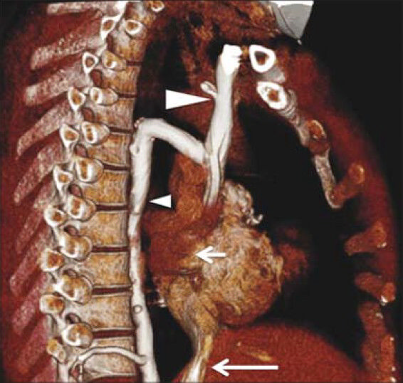
- 18-year-old male with complex congenial heart disease had attacks of ventricular tachycardia and oxygen saturation levels that showed discrepancy between the upper and the lower extremities, later diagnosed with a rare collateral vein draining from the left subclavian vein to the great cardiac vein. Three-dimensional MDCT of the heart and thorax demonstrates extraordinary dilated azygous vein (small arrowhead) draining into the superior vena cava (large arrow head), and the inferior vena cava (large arrow) draining directly into the right atrium (small arrow).
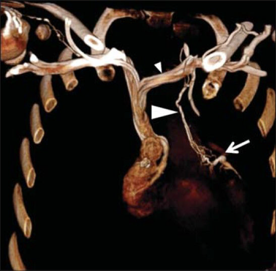
- 18-year-old male with complex congenial heart disease had attacks of ventricular tachycardia and oxygen saturation levels that showed discrepancy between the upper and the lower extremities, later diagnosed with a rare collateral vein draining from the left subclavian vein to the great cardiac vein. Three-dimensional MDCT of the heart and thorax demonstrates the collateral vein (large arrowhead) from the left subclavian vein (small arrowhead) draining into the great cardiac vein (arrow).
MDCT venography technique
Bilateral dual injection MDCT venography was performed using a 256-MDCT scanner (Philips iCT; Philips Healthcare, Clevland Ohio, USA) with the patient positioned supine with arms by his side. Anterior-posterior and lateral scout scans were performed, with a scan range from the apex of the chest to the costophrenic angle. The scan parameters were: Detector width 256 × 0.625 mm; pitch 0.9; rotation time 0.4 s; exposure factors 100 kVp, 200 mA, with z-axis modulation; and scanning time of 2.6 s. A caudocranial scan direction was employed.
Contrast bolus geometry
Bolus geometry is the pattern of enhancement, after intravascular injection of contrast material, measured in a region of interest (ROI), plotted on a time (s)/attenuation Hounsfield units (HU) curve. We employed the test bolus technique where one ROI was plotted inside the superior vena cava and another in the pulmonary trunk. A small amount of contrast material was (10 ml) injected at the same rate as the main bolus using a power injector, where the initial rate was 3 ml/s which was then decreased exponentially to 2.2 ml/s over the duration of the injection using bolus shaping software. This ROI assessed the time to peak (TTP) and determined the arteriovenous circulation time for thoracic vasculature [Figure 3].

- 18-year-old male with complex congenial heart disease had attacks of ventricular tachycardia and oxygen saturation levels that showed discrepancy between the upper and the lower extremities, later diagnosed with a rare collateral vein draining from the left subclavian vein to the great cardiac vein. Determination of contrast bolus transit time using test bolus injection shows that the region of interest is placed inside the superior vena cava (circle A) and in the pulmonary trunk (circle B). Resulting enhancement curves display the time needed to reach the peak of maximum contrast enhancement for test bolus.
Contrast medium administration
Contrast material was injected with an automated dual barrel power injector (Optivantage; Covidien, Cincinnati, OH, USA) via a 20-gauge venous catheter in each arm (right and left sides). Bilateral antecubital venous access was used in this study because it provides uniform contrast distribution into the heart for the contrast material to pass through the venous system with the least amount of dilution, promoting good image quality during computed tomography angiography (CTA) at reduced contrast volumes.[3] Bilateral simultaneous injection occurred at 3 ml/s, employing a 20:80 contrast to saline ratio mix in each syringe. The volume of contrast used totaled to 40 ml (this included 20 ml of contrast and 80 ml of saline in each syringe).
DISCUSSION
Identification of the components of the cardiac congenital anomalies is vital for devising a management plan. Echocardiography and angiography are the current modalities of choice for evaluating congenital heart diseases. However, there has been an increased role of MDCT as a superior imaging modality used prior to surgical intervention for providing detailed anatomical and functional information of venous anomalies with 2D-reformatted and 3D-reconstructed images.[4] This patient required an assessment of the systemic venous return to the heart, especially to the left superior intercostal vein, collateral vein draining into the coronary sinus, and the great cardiac vein. Optimal and diagnostic images are vital in understanding patients’ anatomy and cardiovascular hemodynamics. Additionally, it is necessary to take into consideration the contrast medium administration and parameters that affect bolus geometry as it needs to be carefully configured to match the venous enhancement pattern of complex angioarchitecture during MDCT venography.[56789]
There are limited studies on CT direct venography for evaluating thoracic venous return. However, one study conducted by Kim et al., using CT venography in the diagnosis of benign thoracic central venous obstruction, employed bilateral upper extremity injection with a total volume of 200 ml of contrast medium and scan delay of 20-40 s.[10] However, in our case, we employed a reduced amount of contrast medium (40 ml in total) by using the test bolus technique at the level of the superior vena cava for optimal enhancement.
CONCLUSION
Evaluation of complex venous angioarchitecture in patients with vascular anomalies has considerably improved with the use of MDCT direct venography when injecting contrast media bilaterally and simultaneously via the upper extremity.
ACKNOWLEDGMENT
The authors would like to thank Dr. Ibrahim Al-Sheikh Deeb, Dr. Ali Haydar, and Professor Mukbil Hournai for their contribution to this case study.
Available FREE in open access from: http://www.clinicalimagingscience.org/text.asp?2014/4/1/58/143425
Source of Support: Nil
Conflict of Interest: None declared.
REFERENCES
- Three-dimensional helical CT of pulmonary arteries in infants and children with congenital heart disease. AJR Am J Roentgenol. 1999;173:109-15.
- [Google Scholar]
- Role of CT in the evaluation of congenital cardiovascular disease in children. AJR Am J Roentgenol. 2009;192:1219-31.
- [Google Scholar]
- Effects of right- versus left-arm injections of contrast material on computed tomography of the head and neck. J Comput Assist Tomogr. 2007;31:677-81.
- [Google Scholar]
- Contrast medium administration and parameters affecting bolus geometry in multidetector computed tomography angiography: An overview. JMIRS. 2011;42:113-7.
- [Google Scholar]
- Caudocranial scan direction and patient-specific injection protocols optimize ECG-gated and non-gated thoracic CTA. J Comput Assist Tomogr. 2013;37:725-31.
- [Google Scholar]
- Multidetector computed tomography in the evaluation of cirsoid aneurysm of the scalp - A manifestation of trauma. Clin Imaging. 2013;37:558-60.
- [Google Scholar]
- MDCT angiography of the major congenital anomalies of the extracranial arteries: Pictorial review. J Med Imaging Radiat Oncol. 2013;57:321-8.
- [Google Scholar]
- An optimised patient-specific approach to administration of contrast agent for CT pulmonary angiography. Eur Radiol. 2013;23:3205-12.
- [Google Scholar]
- Role of CT venography in the diagnosis and treatment of benign thoracic central venous obstruction. Korean J Radiol. 2003;4:146-52.
- [Google Scholar]


