
Translate this page into:
Liver Perivascular Epithelioid Cell Tumor with an Unusual Location: Diagnostic Characteristics with Multidetector Computed Tomography and Magnetic Resonance Imaging
Address for correspondence: Assoc. Prof. Koray Hekimoglu, Department of Radiology, School of Medicine, Baskent University, Fevzi Cakmak Avenue, 10. Street Nu: 45 Bahcelievler, Ankara 06490, Turkey. E-mail: korayhekim@yahoo.com.tr
-
Received: ,
Accepted: ,
This is an open access article distributed under the terms of the Creative Commons Attribution-NonCommercial-ShareAlike 3.0 License, which allows others to remix, tweak, and build upon the work non-commercially, as long as the author is credited and the new creations are licensed under the identical terms.
This article was originally published by Medknow Publications & Media Pvt Ltd and was migrated to Scientific Scholar after the change of Publisher.
Abstract
Primary perivascular epithelioid cell tumor (PEComa) of the liver is a very rare tumor that originates from mesenchyma. Gastrointestinal tract with perivascular distribution is the most common anatomic sites of these tumors. Only few cases of hepatic PEComa have been described so far. Malignant PEComas exhibit aggressive behavior with poor prognosis, making early diagnosis crucial. Hereby, we report a 79-year-old female with unusually located mass in the liver. A partial curative hepatectomy has been done, and PEComa was diagnosed histopathologically. No evidence of recurrence was observed during the 6-month follow-up.
Keywords
Computed tomography
liver
magnetic resonance imaging
perivascular epithelioid cell tumor


INTRODUCTION
Perivascular epithelioid cell tumors (PEComas) are seldom group of mesenchymal tumors, and etiology of these tumors remains uncertain.[1] PEComas are known for their specific perivascular location; they originates from perivascular epithelioid cells. These lesions are generally benign natüre and often curable with surgery. The most common locations of PEComa are the ovaries, uterus, rectum, bladder, abdominal wall, heart, and pancreas.[2] Another important site of this tumor – that we emphasized – is in the liver. Although PEComas are commonly silent, gastrointestinal PEComas commonly present with nonspecific signs. First signs including abdominal pain, tenderness, melena, anemia, impaction, or obstruction due to enlargement of tumor volume.
A little portion of PEComas reveals aggressive form and generally have a female predominance with rapid local invasion and distant metastases.[3] In this case report, mortal course is inevitable if the process is rapid and curative treatment is inadequate.
Today, literatüre on multimodal imaging characteristics of PEComa is not sufficient. With increasing awareness of this entity, it is very important to be familiar with the evaluation of PEComas across different imaging modalities as multidetector computed tomography (MDCT) and magnetic resonance imaging (MRI) with fast multiplanar imaging techniques.
Hereby, we demonstrated the appearance of hepatic PEComa on unexpected location of liver in one case.
CASE REPORT
A 79-year-old female who was previously healthy admitted to our hospital with a 1 year history of constant dull right upper quadrant pain and tenderness. All laboratory tests including tumor markers were in normal limits. There was not any story of hepatitis or chronic liver disease. Abdominal ultrasound revealed a hypoechoic solid lesion that located in the right and caudate lobe of liver with irregular border. MDCT examination reveals a liver mass with 52 mm × 43 mm diameters located in segment VIII and caudate lobe of the liver [Figure 1]. The lesion showed heterogeneous enhancement in arterial phase with hypointense central area also. The mass become hypodense to isodense to adjacent parenchyma on delayed venous phase. The region of nonenhancing hypointense area was likely to represent central necrosis, which was frequently seen in malign PEComas. The patient underwent MRI, after first assessment with MDCT for better evaluation of vascular invasion. In MRI, the tumor had intermediate isointense signal on T1-weighted images and heterogeneously hyperintense on T2-weighted images relative to the liver parenchyma [Figure 2]. There was not seen any fat component in the lesion. On gadolinium (Gd)-enhanced images, the mass showed significant and heterogeneous enhancement. Nonenhancing hypointense area in tumor center representing necrosis. Inferior vena cava (IVC) invasion with tumor thrombus into the lümen is also demonstrated, especially coronal Gd-enhanced images [Figure 3]. There was also marked diffusion restriction of lesion on diffusion-weighted images as totally hypointense signal in apparent diffusion coefficient maps [Figure 4].

- Imaging of a hepatic perivascular epithelioid cell tumor. (a) Contrast-enhanced arterial phase of computed tomography reveals a poorly defined mass with marked enhancement and inferior vena cava invasion. (b) Coronal reconstruction of venous phase contrast-enhanced computed tomography shows relatively washout of mass with unclear borders. Tumor thrombus in inferior vena cava is also seen clearly in coronal multidetector computed tomography image.
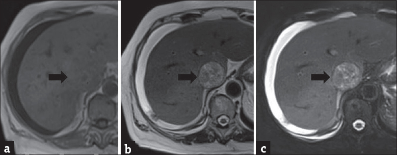
- Axial T1-weighted (a), T2-weighted (b), and fat-suppressed T2-weighted (c) magnetic resonance imaging images demonstrate T1-isointense, T2 and fat-suppressed T2-hyperintense mass component of liver perivascular epithelioid cell tumor in vena cava inferior.
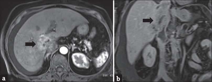
- Postgadolinium fat-suppressed axial arterial phase (a), and coronal venous phase images demonstrate a large liver perivascular epithelioid cell tumor that marked heterogeneous enhancement in arterial phase, and washout in venous phase with prominent invasion of inferior vena cava. Tumor thrombus is markedly seen in late venous phase (b).
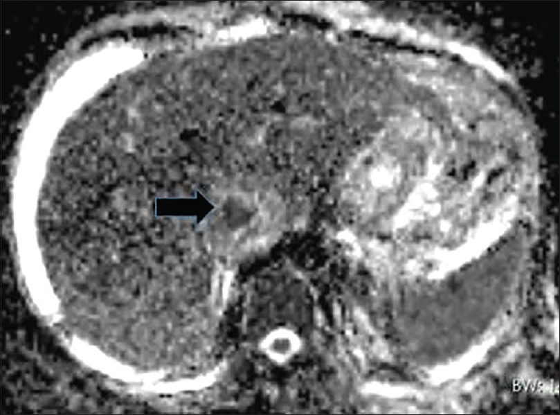
- Diffusion-weighted image of perivascular epithelioid cell tumor (black arrow). Marked diffusion restriction of malign perivascular epithelioid cell tumor is seen as hypointense signal change on apparent diffusion coefficient map.
Calcification and/or hemorrhage were not detected in the lesion. Metastasis in other systems was also not found. The patient consented to tumor biopsy after radiologic assessment. The patient underwent biopsy of the liver mass, and pathology revealed a hepatic perivascular epithelioid cell tumor or “PEComa.” Histologic findings were a mixture of large epithelioid cells and clear-to-eosinophilic granular cytoplasm [Figure 5]. There was severe nuclear atypia with large nucleoli and increased mitotic activity detected microscopically. However, immunochemistry study indicated that tumor cells were positive for human melanoma black (HMB)-45 marker. Following the surgery, the patient has not shown any evidence of recurrence for 6 months.
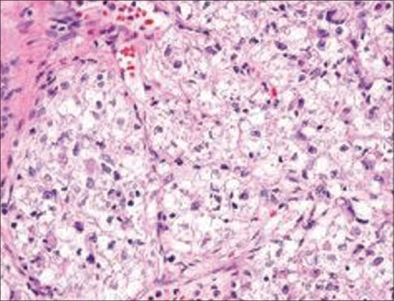
- Histopathologic findings of malign perivascular epithelioid cell tumor; photomicrograph (H and E, ×400) shows specimen is composed of nests of large epithelioid cells with clear-to-granular eosinophilic cytoplasm. There is also seen severe nuclear atypia that indicates of malign tumor.
DISCUSSION
PEComa was first proposed in 1992 by Bonetti et al. and also classified by the World Health Organization in 2002 as a mesenchymal tumor.[4] However, perivascular epithelioid cells were first defined by Apitz in 1944. PEComas are tumors that consist of epithelioid cells with histologically and immunohistochemically specific markers for melanocytes and smooth muscles.[5] This tumor had extensive family with various cytologic subtypes that include classic and epithelioid angiomyolipoma, clear cell tumor of the lung, lymphangioleiomyomatosis, clear cell tumors of the retroperitoneum, rectum, heart, pancreas, and uterus, and also myomelanocytic clear cell tumor of the ligamentum teres in liver.[6]
PEComas are predominantly seen in women in the first three decades (3–29 years) of life. These tumors are mostly asymptomatic in early stages. However, it may cause vaguely pain once they grow to moderately large size. Liver PEComas have a tendency to locate at falciform ligament/ligamentum teres. Folpe et al. reported seven cases with liver PEComas located within or abutting near the falciform ligament or ligamentum teres.[7] PEComas may be located in another place of liver rarely as seen in this case report. Besides, in this case, the mass was located at the medial aspect of the liver with the invasion of IVC. In literature, it was not concluded any IVC invasion of PEComa until today.
Hepatic PEComas usually exhibit distinct margins. In general, PEComas have been tend to exhibit benign behavior in the majority of the cases. However, several malignant PEComas have been reported.[356] It was concluded that tumor was >5 cm, and concomitant necrosis was criteria for malignancy.8 In another study, Tan and Xiao detected malign PEComas range accepted from 2.5 to 8.5 cm with a mean of 4 cm.[9]
Ultrasound, computed tomography (CT) and MRI are not sufficiently sensitive to enable exact diagnosis of PEComa because of its nonspecific radiologic characteristics. Furthermore, distinct criteria between benign and malign types of PEComas have not been defined in most of the studies until today.[1011]
CT and MRI studies should be made with contrast enhanced for exact evaluation of liver PEComas. Typically, lesion reveals heterogeneous enhancement on arterial and early portal phase of contrast-enhanced CT and MRI.[6] The imaging characteristics of hepatic PEComa may commonly mimic malignant liver tumors such as hepatocellular carcinoma or metastases. Hypervascularity and also arteriovenous connections could be responsible for misdiagnosis in some cases. On the other hand, chronic liver disease is cooccurrence with liver PEComas in the elderly. If the liver parenchyma is normal appearance, hepatocellular carcinoma should not be considered, firstly. Literature reviews suggest that hepatic PEComas were detected in livers without a background of cirrhosis or hepatitis, also.[9] Nonenhancement areas in the mass with low attenuation generally show internal necrosis, which is frequently due to the rapid growth of PEComas.
Hepatic PEComas are mostly isointense to mildly hypointense to muscle on T1-weighted images on MRI. If the presence hemorrhagic components into the mass, it could be seen as hyperintense on fat-suppressed T1-weighted images. Heterogeneous hyperintense signal is characteristic sign on T2-weighted images for PEComas, especially for malign ones. Besides, some regions in the lesion with T2-shortening brightness could be seen due to internal protein component or hemorrhage.[10] Tumor margins with well demarcated are better seen on T2 images. Contrast-enhanced sequences always should be added to MR studies in PEComas.
Enhancement characteristics of PEComas are very variable; a recent study concluded that PEComas tend to show intense enhancement.[11] However, PEComas may show persistent enhancement in late phases in some cases, which gives rise to mistake as benign masses such as focal nodular hyperplasia and hemangioma.[69]
Exact diagnostic type of PEComa depends on the pathology and also immunohistochemistry. PEComa cells show similarity to epithelioid cells with dilated vascular channels and contain eosinophilic cytoplasms. Nevertheless, all of the PEComas nearly are diagnosed with positive melanocytic markers and smooth muscle markers in studies.[412] In this case report, the tumor was positive for HMB-45 marker also.
Today, surgery with adequate margins remains the gold standard for treatment of PEComas, especially in malignant ones.
CONCLUSION
PEComa is detected increasingly in liver. Dynamic and multiplanar imaging should be done with MDCT and MRI at invasive liver lesions. If an invasive lesion was found in liver without a background of hepatitis or cirrhosis, liver PEComa should be considered in differential diagnosis. The exact diagnosis of PEComa depends on the pathologic observations basically. Surgery appears to be necessary for a total cure. These tumors show benign behavior mostly, and the prognosis seems good after surgery.
Financial support and sponsorship
Nil.
Conflicts of interest
There are no conflicts of interest.
Available FREE in open access from: http://www.clinicalimagingscience.org/text.asp?2017/7/1/36/215048
REFERENCES
- Perivascular epithelioid cell tumor of the liver: A report of two cases and review of the literature. World J Gastroenterol. 2007;13:5537-9.
- [Google Scholar]
- Malignant perivascular epithelioid cell tumor (PEComa) of the femur: A case report and literature review. Diagn Pathol. 2015;10:54.
- [Google Scholar]
- Management of perivascular epithelioid cell tumor of the liver: A case report and review of the literature. Oncol Lett. 2014;7:148-152.
- [Google Scholar]
- Malignant hepatic perivascular epithelioid cell tumor (PEComa) - Case report and a brief review. J Egypt Natl Canc Inst. 2015;27:239-42.
- [Google Scholar]
- Update on the imaging of malignant perivascular epithelioid cell tumors (PEComas) Abdom Radiol (NY). 2016;41:368-76.
- [Google Scholar]
- Clear cell myomelanocytic tumor of the falciform ligament/ligamentum teres: A novel member of the perivascular epithelioid clear cell family of tumors with a predilection for children and young adults. Am J Surg Pathol. 2000;24:1239-46.
- [Google Scholar]
- “Malignant” perivascular epithelioid cell neoplasm: Risk stratification and treatment strategies. Sarcoma 2012 2012:541626-38.
- [Google Scholar]
- Hepatic perivascular epithelioid cell tumor (PEComa): Dynamic CT, MRI, ultrasonography, and pathologic features – Analysis of 7 cases and review of the literature. Abdom Imaging. 2012;37:781-7.
- [Google Scholar]
- PEComa: A perivascular epithelioid cell tumor in the liver-A case report and review of the literatüre. Case Rep Med 2013 2013:904126.
- [Google Scholar]
- Imaging features of primary and metastatic malignant perivascular epithelioid cell tumors. AJR Am J Roentgenol. 2014;202:252-8.
- [Google Scholar]
- Primary perivascular epithelioid cell tumor of the liver not related to hepatic ligaments: Hepatic PEComa as an emerging entity. Histol Histopathol. 2008;23:1185-93.
- [Google Scholar]


