Translate this page into:
Isolated Humeral Metastasis in Uterine Cervical Cancer: A Rare Entity
Address for correspondence: Dr. Ramin Pourghorban, Department of Radiology, Shohada-e-Tajrish Hospital, Shahid Beheshti University of Medical Sciences, Tehran, Zip code-1989934148, Iran. E-mail: ramin_p2005@yahoo.com
-
Received: ,
Accepted: ,
This is an open-access article distributed under the terms of the Creative Commons Attribution License, which permits unrestricted use, distribution, and reproduction in any medium, provided the original author and source are credited.
This article was originally published by Medknow Publications & Media Pvt Ltd and was migrated to Scientific Scholar after the change of Publisher.
Abstract
Bone metastasis in cancer of uterine cervix, especially in the form of isolated bone involvement is a rare manifestation. Herein, we report the first case of isolated humeral metastasis in a known case of locally advanced cervical cancer. A fifty-six-year old female presented with International Federation of Gynecology and Obstetrics (FIGO) Stage IV A squamous cell carcinoma of uterine cervix. She was treated with a combination of radiation and chemotherapy and then total abdominal hysterectomy with bilateral salpingo-oophorectomy. Seven months later, she developed an isolated lytic lesion in the left humerus, which turned out to be a bone metastatic lesion.
Keywords
Bone metastasis
cervical cancer
humerus
INTRODUCTION

Cervical cancer is the third most common gynecologic malignancy. About 80%-90% of cervical carcinomas are of squamous cell origin. Cancer of uterine cervix arises from the squamo-columnar junction of the cervix and may develop from precursor lesions in preclinical stages, including carcinoma in situ, cervical intraepithelial neoplasia, and severe dysplasia. Bone metastasis from carcinoma cervix is rare, especially in isolated bone involvement of the distal appendicular skeleton. Thanapprapasr et al.,[1] reported 51 (1.1%) patients with bone metastasis among 4620 cervical cancer patients. Fagundes et al.,[2] reported that in a retrospective analysis of 1211 patients with invasive carcinoma of the uterine cervix, incidence of distant metastases was 26% in Stage IIB. They usually occur in the spine and pelvis. There are a few reported cases of isolated localized metastasis to fibula, patella, and skull arising from cancer of uterine cervix in the literature.[3–5] To our knowledge, this is the first case of uterine cervix cancer with localized metastasis to the humerus.
CASE REPORT
A 56-year old post-menopausal female, gravida 5, para 3, was admitted to our hospital, with a 3-month history of abnormal vaginal bleeding. She underwent colposcopy-guided biopsy and the diagnosis was moderately differentiated invasive squamous cell carcinoma of uterine cervix, mixed small and large cell non-keratinizing variants. Work-up assessments including chest X-ray, abdomino-pelvic ultrasound exam, and contrast-enhanced pelvic MRI were done. The primary tumor, measuring 130 mm in its largest diameter, was located in the cervix and invasion of myometrium and the parametrial region were also revealed; nevertheless, there was no evidence of distant metastasis or lymph node enlargement. According to the imaging findings, the stage of the tumor was estimated to be at least IIB (T2b, N0, M0) and the patient underwent combined radio-chemotherapy followed by total abdominal hysterectomy and bilateral salpingo-oophorectomy, according to our hospital protocol. The pathology confirmed the above mentioned findings as well as tumoral involvement of left adenex; whereas, the right ovary was unremarkable. Rectosigmoid and bladder wall biopsies also showed metastatic involvement. In addition, peritoneal washing result for malignancy was positive. Thereby, the accurate stage of the disease was defined as IVA (T4, N0, M0).
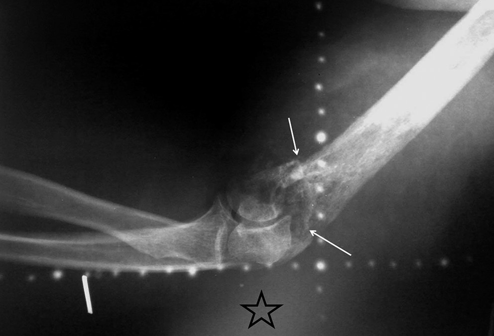
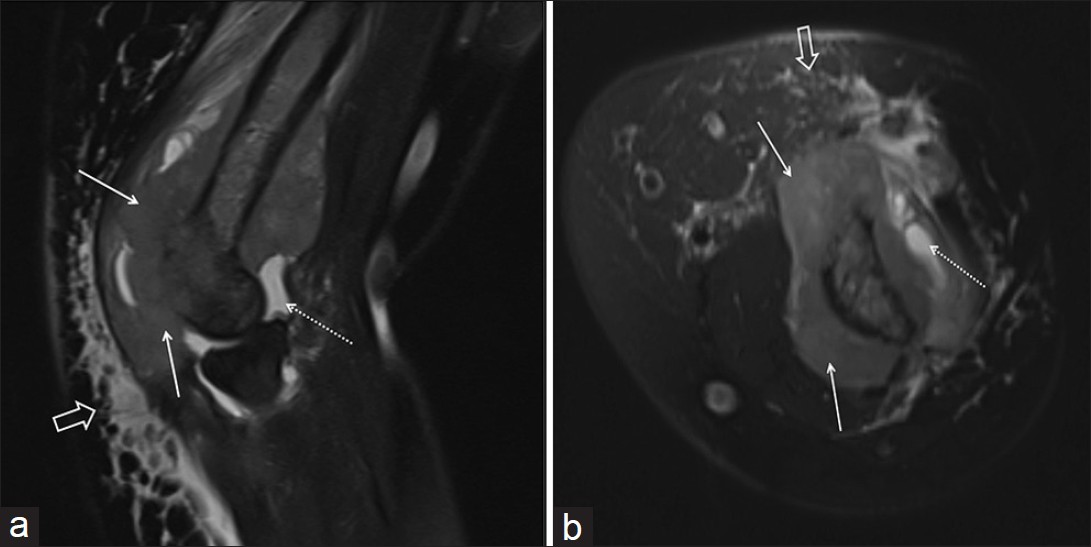
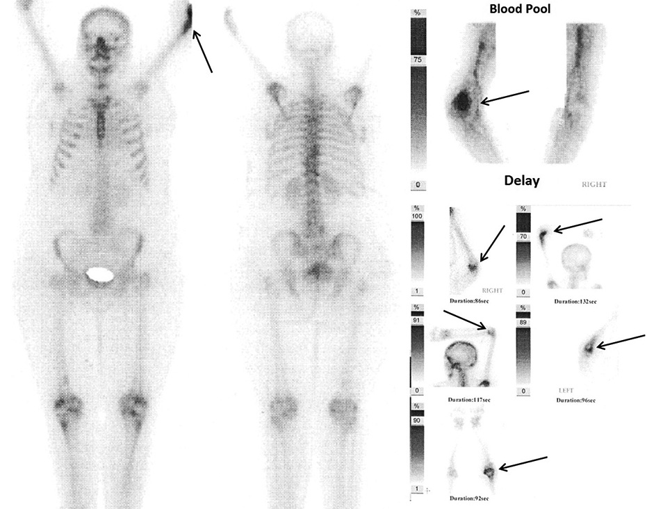
Seven months after the treatment, she developed left elbow pain. Physical exam revealed decreased range of motion accompanied by soft tissue swelling in left elbow. Thereafter, plain X-ray, MRI of left upper extremity, and whole body bone scan were performed. In plain radiograph [Figure 1] a destructive lytic lesion with pathologic fracture was found in the distal humerus. In the MRI exam [Figure 2] a soft tissue mass with T1 hypo-signal and T2 intermediate-signal intensities was seen surrounding the distal part of the left humerus associated with destruction of bone cortex. Isotopic bone scan [Figure 3] showed isolated increased tracer concentration at the distal part of the left humerus whereas no evidence of skeletal metastasis was found elsewhere. The biopsy from the bone lesion was taken and pathological examination of resected specimen confirmed metastatic squamous cell carcinoma [Figure 4].

- Plain radiograph before initiation of radiotherapy shows an irregular and poorly defined destructive lytic lesion with no periosteal reaction in distal humerus associated with pathologic fracture (white arrows) and abnormal adjacent soft tissue density (star).

- (a) Sagittal and (b) Axial fat suppressed T2-weighted images demonstrate soft tissue mass (white arrows) surrounding the left humerus with destruction of bone cortex and replacement of the involved bone marrow with non-homogenous intermediate signal intensity compared to adjacent bone marrow. Also noted are joint effusion (dashed arrows) accompanied by subcutaneous edema (open arrows).

- Tc99 MDP isotopic bone scan demonstrates isolated increased tracer concentration at the distal of left humerus (arrows) which is also persistent in delayed images (right lower corner). Scintigraphic evidence of skeletal metastasis was not found elsewhere.

- Section shows a neoplasm composed of pleomorphic, large nonkeratinizing and high nucleus to cytoplasm ratio cells with marked nucleoli arranged in sheet formations which infiltrate soft tissue and bony trabeculi (arrows). Also noted are some foci of necrosis. These findings are consistent with poorly differentiated metastatic squamous cell carcinoma.
DISCUSSION
In a few autopsy series, the prevalence of bone metastases in the setting of recurrent cervical carcinoma was reported in 15%-29%.[6] The most common presenting symptom in bone metastasis is pain. The vertebral bodies are the most common site of osseous metastasis, followed by the pelvis, ribs, and extremities. Bone metastasis in squamous cell carcinoma of the cervix may occur by lymphogenous or hematogenous spread or by direct extension from adjacent lymph nodes.[6] Blythe et al.,[7] found that the most common mechanism of bone involvement was by direct extension of neoplasm from para-aortic nodes into the adjacent vertebral bodies. In contrast, metastasis to distant long bones and calvarium is supposed to be hematogenous.[8] Matsuyama and colleagues,[9] evaluated the rates of bone recurrence in 713 patients with cancer of the uterine cervix and reported it as 4.0% in stage I, 6.6% in stage II, 8.0% in stage III, and 22.9% in stage IV, respectively. Metastasis to skull, clavicle, ribs, scapula, sternum, tarsal, metatarsal and involvement of both tibia and fibula and isolated metastasis of tibia, fibula and femur has been reported in literature, whereas isolated metastasis to humerus from cancer of uterine cervix has not been previously documented.[34710] Therefore, to the best of knowledge, this is the first reported case of isolated metastasis to humerus from cancer of uterine cervix.
Plain radiograph, bone CT are useful modalities for evaluation of metastatic bone disease. MRI depicts the soft tissue extension to a better advantage. In addition, bone scan is also a useful diagnostic tool for detection of distant metastasis and to differentiate the isolated distant metastasis from diffuse metastasis. Due to short life expectancy of patients with bone metastasis, the treatment is usually directed toward preservation of quality of life. Till now, there is no specific widely accepted guideline for the treatment of patients with bone involvement; nevertheless, in case of isolated and resectable bone metastasis, complete surgical excision followed by palliative chemo-radiotherapy is recommended.[4] Given the poor prognosis of bone metastasis, the benefit of the mentioned approach is still questionable and also depends on the patients’ compliance. As in our case, the patient refused the offer of surgical excision and underwent palliative radiation and palliative therapy.
CONCLUSION
Bone metastasis from carcinoma cervix is infrequent and causes dismal prognosis in locally advanced cervical cancer. Whole body bone scan can detect distal bone metastasis and distinguish isolated bone metastasis from diffuse metastasis; whereas, MRI can clearly show tumor extension into adjacent soft tissue. There is no widely-accepted approach for the treatment of patients with bone involvement. Nevertheless, bone metastasis brings dismal outcome; hence, the treatment is usually directed toward maintaining the quality of life of the patients.
Available FREE in open access from: http://www.clinicalimagingscience.org/text.asp?2012/2/1/80/105137
Source of Support: Nil
Conflict of Interest: None declared.
REFERENCES
- Bone metastasis in cervical cancer patients over a 10-year period. Int J Gynecol Cancer. 2010;20:373-8.
- [Google Scholar]
- Distant metastases after irradiation alone in carcinoma of the uterine cervix. Int J Radiat Oncol Biol Phys. 1992;24:197-204.
- [Google Scholar]
- Femur metastasis in carcinoma of the uterine cervix: A rare entity. Arch Gynecol Obstet. 2010;281:963-5.
- [Google Scholar]
- Carcinoma of uterine cervix with isolated metastasis to fibula and its unusual behavior: Report of a case and review of literature. J Cancer Res Ther. 2006;2:79-81.
- [Google Scholar]
- Carcinoma of the cervix with patellar metastases: Case report. Am J Cancer. 1937;29:122-4.
- [Google Scholar]
- Distant metastases in squamous-cell carcinoma of the uterine cervix. Radiology. 1967;88:961-6.
- [Google Scholar]


