
Translate this page into:
Honda Sign On 18-FDG PET/CT in a Case of Lymphoma Leading to Incidental Detection of Sacral Insufficiency Fracture
Address for correspondence: Dr. Prathamesh Joshi, Department of Nuclear Medicine and PET-CT, Jaslok Hospital and Research Centre, Worli, Mumbai – 400 026, India. E-mail: drprathamj@gmail.com
-
Received: ,
Accepted: ,
This is an open-access article distributed under the terms of the Creative Commons Attribution License, which permits unrestricted use, distribution, and reproduction in any medium, provided the original author and source are credited.
This article was originally published by Medknow Publications & Media Pvt Ltd and was migrated to Scientific Scholar after the change of Publisher.
Abstract
Sacral insufficiency fracture (SIF) is an important and treatable cause of low back pain in at-risk groups and the elderly. We report rare demonstration of ‘Honda sign’ in fluoro-deoxy-glucose positron emission tomography-computed tomography (FDG PET-CT) in a case of lymphoma, which led to incidental diagnosis of SIF. Honda sign, which is classically described in bone scans in cases of SIF, was found in FDG PET-CT in our case. This suggests SIF should be suspected when similar FDG uptake pattern is encountered and may help in early detection and management of SIF.
Keywords
Bone scan
fluorodeoxyglucose
honda sign
positron emission tomography-computed tomography
sacral insufficiency fracture

INTRODUCTION

Fluoro-Deoxy-Glucose Positron Emission Tomography-Computed Tomography (FDG PET-CT), often regarded as the “one-stop shop” for diagnosing many malignancies, provides co-registered structural and metabolical images, allowing for accurate localization of disease sites. In malignancies, the procedure has become essential in staging of the disease, monitoring response to treatment, planning and choosing appropriate therapies, detecting recurrence, and providing accurate prognoses. However, non-specific FDG accumulation observed at sites of inflammation, during PET imaging of patients with cancer, is a known phenomenon and it has evolved into a promising imaging technique to examine, diagnose, and manage inflammatory disorders.[1]
We report FDG PET-CT findings in a case of lymphoma where increased FDG uptake in the sacroiliac region led to incidental diagnosis of sacral insufficiency fracture (SIF).
CASE REPORT
A 63-year-old woman, a known case of non-Hodgkin's lymphoma, underwent whole body FDG PET-CT scan for surveillance. She had Stage I disease involving the left axillary lymph nodes at the time of diagnosis and had completed chemotherapy a year ago. She was in remission since then. The patient had complained of lumbago for the past 4 months. The FDG-PET scan (Maximum Intensity Projection- Figure 1) showed vertical linear FDG uptake in the medial to bilateral sacroiliac joint and horizontal uptake connecting the vertical lines (H-shaped uptake) in the sacral region. This ‘H’-shaped increased FDG uptake resembled the typical Honda/Butterfly sign, classically described in bone scans in cases of SIF. There was no evidence of abnormal FDG uptake in left axillary region (site of primary disease) or anywhere else in the body. The axial and coronal maximum intensity projection (MIP) CT images [Figure 2a and 2b] show lucent fracture line (arrow) with breach in the anterior cortex of right sacral ala. The fracture line is away from the right sacral foramina and medial to the sacroiliac joint. There was no history of trauma to sacroiliac region or any history of a fall. We concluded the scan abnormality as SIF.
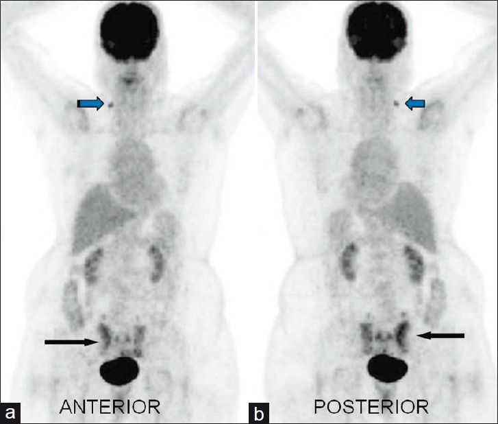
- The F-18 FDG PET-CT (maximum intensity projection image) (a) Anterior and (b) posterior show focal low-grade tracer uptake in right level IV cervical lymph node (nonspecific) blue arrow, and vertical linear FDG uptake medial to bilateral sacroiliac joint and horizontal uptake which connects vertical line (H-shaped/ Butterfly shaped) (black arrow) in the sacral region. Standardized uptake maximum value of sacral uptake based on body surface area was 5.8 gm/ml.
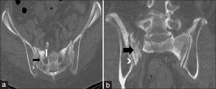
- The (a) axial and (b) coronal Maximum Intensity Projection (MIP) CT images show lucent fracture line (black arrow) with breach in the anterior cortex of right sacral ala. The fracture line is away from the right sacral foramina (white arrow) and medial to the sacroiliac joint (arrowhead).
A correlation with nuclear magnetic resonance (NMR) imaging of sacral spine was performed. Short tau inversion recovery (STIR)-weighted axial and coronal images [Figure 3a and 3b] through the sacrum revealed complete diffuse hyperintensity of the right sacral ala suggestive of bone marrow bruise/edema. There was also mild hyperintensity in the soft tissue anterior to the right sacral ala suggestive of changes related to bone injury. Bilateral SI joints and bilateral iliac bones appeared unremarkable. These features were consistent with SIF. The patient was referred to a rheumatologist for further management and is being treated conservatively.
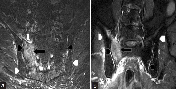
- Nuclear magnetic resonance (NMR) images of sacral spine. Short tau inversion recovery (STIR) weighted (a) axial and (b) coronal images through the sacrum revealed complete diffuse hyperintensity of the right sacral ala (black arrow) suggestive of bone marrow bruise/edema. There is also mild hyperintensity in the soft tissue anterior (white arrow) to the right sacral ala suggestive of changes related to bone injury. Bilateral SI joints (black arrowhead) and bilateral iliac bones (white arrowhead) appear unremarkable. These features were suggestive of sacral insufficiency fracture.
DISCUSSION
SIF is a kind of stress fracture, which occurs due to the effect of normal or physiological stress on weakened bone with decreased elastic resistance.[2] The clinical presentation is variable, although there is usually severe lower back pain, often exacerbated by movement and radiating to the leg or groin, with no history of trauma. There are multiple risk factors for insufficiency fractures, the most common being osteoporosis and then rheumatoid arthritis, prolonged corticosteroid treatment and pelvic irradiation.[3–6]
Elderly patients often present with lower back pain and related symptoms. There are many possible diagnoses including malignancy and osteomyelitis. Insufficiency fractures involving the sacrum are an important and treatable cause of severe back pain, but are under-reported.[3–5] There is also potential for misdiagnosis as there is often a history of prior malignancy in the at-risk patient.[346]
Bone scintigraphy is one of the most sensitive technique for detecting SIF, with the H-shaped or Butterfly pattern of uptake regarded as being diagnostic.[78] The classic “H” pattern is produced when there are fractures of both sacral alae and a horizontal component involving the sacral body.[3] In a meta-analysis, the classical H-sign was documented in 40% patients of SIF.[5]
Our case represents rare demonstration of ‘Honda sign’ in FDG PET-CT. There are very few case reports in literature describing, incidental detection of SIF in FDG PET-CT of the patients undergoing the scan for evaluation of malignancy.[910] The mechanism of increased FDG uptake in the fracture site is thought to be due to the migration of activated inflammatory cells.[11] Knowledge of such uptake is important in reporting FDG PET-CT, to avoid misinterpretation of the uptake as metastatic bone involvement, resulting in false positive scan. CT findings and NMR, bone scan correlation are valuable tools to confirm the cause of FDG uptake in similar case scenarios.
CONCLUSIONS
Our case represents a rare demonstration of ‘Honda Sign’ on FDG PET-CT scan in a patient leading to the diagnosis of SIF. The case suggests SIF should be suspected when similar FDG uptake pattern is encountered. This may help in the early detection and management of SIF.
Source of Support: Nil
Conflict of Interest: None declared.
Available FREE in open access from: http://www.clinicalimagingscience.org/text.asp?2012/2/1/29/96544
REFERENCES
- Unparalleled contribution of 18F-FDG PET to medicine over 3 decades. J Nucl Med. 2008;49:17N-21N.
- [Google Scholar]
- Sacral insufficiency fractures: A report of two cases and a review of the literature. J Womens Health Gend Based Med. 2001;10:699-705.
- [Google Scholar]
- Fractures of the sacrum caused by bone insufficiency.Meta-analysis of 508 cases. Presse Med. 1997;26:1568-73.
- [Google Scholar]
- Stress fractures. In: Ferrucci JT, Taveras JM, eds. Radiology. Philadelphia, PA: Lippincott, Williams and Wilkins; 2002.
- [Google Scholar]
- Unsuspected sacral fractures: Detection by radionuclide bone scanning. AJR Am J Roentgenol. 1985;144:337-41.
- [Google Scholar]
- Sacral insufficiency fracture detected by FDG-PET/CT: Report of 2 cases. Ann Nuc Med. 2006;20:445-8.
- [Google Scholar]
- Avoidance of Misinterpretation of an FDG Positive Sacral Insufficiency Fracture Using PET/CT Scans in a Patient With Endometrial Cancer: A Case Report. Clin Nucl Med. 2007;32:779-81.
- [Google Scholar]
- Rapid normalization of osseous FDG uptake following traumatic or surgical fractures. Eur J Nucl Med Mol Imaging. 2003;30:1096-103.
- [Google Scholar]


