
Translate this page into:
Granulomatous mastitis following stereotactic core-needle biopsy: A case report

*Corresponding author: Laura L. Kirk, College of Medicine, University of Kentucky College of Medicine, Lexington, United States. lles222@uky.edu
-
Received: ,
Accepted: ,
How to cite this article: Kirk LL, McDonald RJ, Clark AX. Granulomatous mastitis following stereotactic core-needle biopsy: A case report. J Clin Imaging Sci. 2024;14:40. doi: 10.25259/JCIS_53_2024
Abstract
Idiopathic granulomatous mastitis is a rare, chronic inflammatory disease of the breast of uncertain etiology that can mimic breast cancer. In rare instances, it may emerge secondary to trauma to the breast. We present a case of a 66-year-old woman who initially underwent a benign stereotactic core-needle biopsy of her left breast complicated by a small hematoma which initially remained unchanged mammographically and sonographically for 1 year; then, it enlarged unexpectedly at the 21-month interval follow-up prompting an ultrasound-guided biopsy revealing granulomatous mastitis.
Keywords
Granulomatous mastitis
Breast
Breast cancer
Inflammatory breast disease
Mastitis

INTRODUCTION
Idiopathic granulomatous mastitis (IGM), also known as granulomatous lobular mastitis, is a rare but benign chronic inflammatory disease of the breast that continues to challenge clinicians with its elusive etiology, unpredictable presentations, and diagnostic complexities. While little is known of the etiology of granulomatous mastitis (GM), an autoimmune, infectious, or hypersensitivity reaction are among the most commonly proposed theories.[1] Cases associated with trauma to the epithelium of the mammary ducts are also gaining attention.
GM typically presents in women of childbearing age, 20–40 years old, typically within the first few years postpartum.[2,3] Clinical presentation varies widely, encompassing painless localized masses that grow slowly over time, painful erythematous masses, inflammation, induration, fistula formation, skin ulceration, nipple discharge, and nipple retraction.[3] The diagnosis of GM poses a formidable challenge due to its rarity and varied clinical presentation along with non-specific radiographic findings which vary based on the timing of radiographic evaluation, extent of inflammation, and possibility of prior intervention. It is essential to rule out carcinoma of the breast due to its similar presentation. Definitive diagnosis is based on histological evidence of lobulocentric granulomatous formations within the biopsied breast tissue.[4] Treatment options are not clearly established but include observation, corticosteroids, and immunosuppressants. Often, surgical management is the last resort, although lesions may recur and result in poor esthetic outcomes.[1]
CASE REPORT
A 66-year-old woman, without personal history of breast cancer, presented for her annual screening mammogram which detected a 3 mm group of calcifications in the left breast, at the 12 o’clock position, 3 cm from the nipple, which were characterized as amorphous on diagnostic mammography [Figure 1] and recommended for a stereotactic core-needle biopsy. Post-biopsy, there was a 2 cm hematoma [Figure 2A]. Pathology resulted in benign fibrosis and associated microcalcifications, concordant with imaging findings. On the initial 6-month post-biopsy follow-up, there was a slight expected decrease in size of the hematoma [Figure 2B], which remained unchanged at 1 year follow-up [Figure 2C].
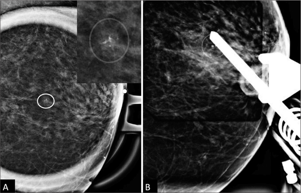
- A 66 year old woman’s screening mammogram with Spot magnification left CC view shows a 3mm group of amorphous calcifications (white circle), with further digital magnification in the top right corner for improved resolution. (A) There is no change in the shape of the calcifications in the magnified ML view (not shown). (B) Postfire stereotactic image shows the needle positioned appropriately at the trough, targeting the calcifications.
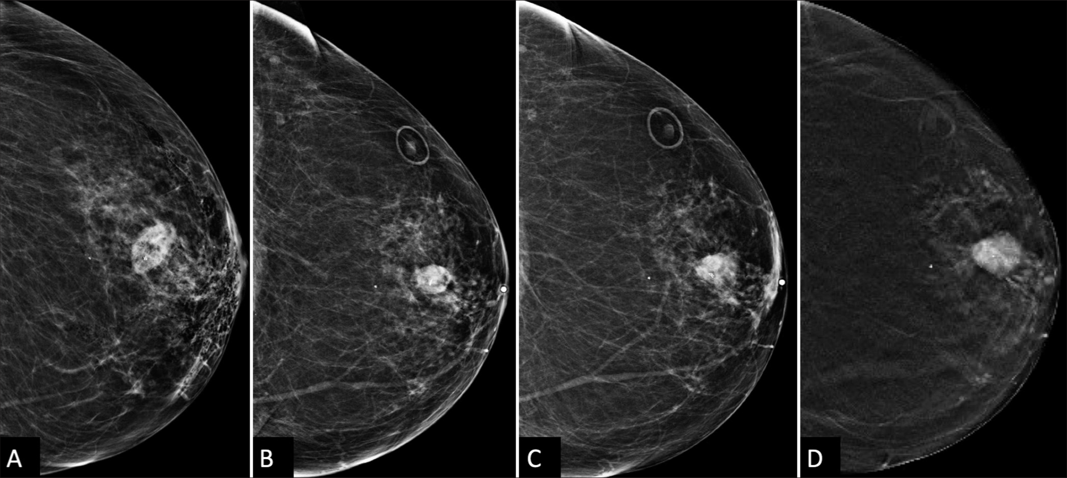
- A 66 year old woman’s Post-biopsy changes in the left breast. (A) Immediate post-biopsy CC view shows a 21 mm × 13 mm hematoma and emphysema in the anterior subareolar region. (B) A follow-up mammogram 9 months later shows an interval decrease in the size of the hematoma to 18 mm × 11 mm (white circle depicting skin marker) (C) Six months after that, the hematoma remained stable to minimally increased, measuring 19 mm × 12 mm (also confirmed on sonography, not shown). (D) At the 21-month follow-up, CC tomosynthesis shows an increase in the size of the hematoma to 21 mm × 14 mm with surrounding distortion or spiculations radiating from the mass, raising suspicion and prompting ultrasound-guided biopsy.
At 21 months post-biopsy, there was interval increased size, density, and subtle new distortion of the mammographic mass known to represent a post-procedural hematoma [Figure 2D]. Targeted left breast ultrasound at the 12 o’clock position, 3 cm confirmed increased size of the hematoma now 20 mm in size [Figure 3A and B]. Given interval enlargement, a subsequent ultrasound-guided core biopsy was performed with partial dissipation of the complex cystic and solid mass and resultant pathology of GM [Figure 4A]. The case was reviewed in multidisciplinary conference, and given patient was not symptomatic, a conservative watchful approach was taken.
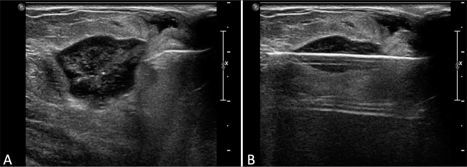
- A 66 year old woman’s Ultrasound-guided biopsy of a complex cystic and solid oval mass. (A) Pre-fire ultrasound image shows the mass, representing a post-biopsy hematoma from a previous stereotactic-guided biopsy 21 months earlier. (B) Post-fire image shows appropriate needle placement within the targeted mass.
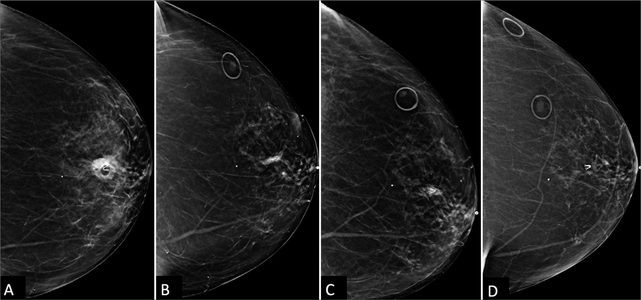
- A 66 year old woman’s Post-ultrasound-guided biopsy changes in the left breast. (A) Immediate post-biopsy CC view shows a new U-shaped clip and a minimally decreased hematoma size of 20 mm × 12 mm (white circle). Pathology confirmed granulomatous mastitis (GM). (B) A follow-up mammogram 6 months later shows near complete resolution of the hematoma (white circle) with conservative observation. However, the patient developed skin cellulitis, indicated by the arrow at the area of focal skin thickening at the biopsy site, which resolved with antibiotics. (C) The hematoma and was not seen on the subsequent annual screening mammogram (white circle). (D) The following screening mammogram shows complete resolution of the prior subtle sub-centimeter asymmetry near the U-shaped clip (white circle), confirming biopsy-proven GM.
At her next clinical visit, the patient reported waxing and waning of skin erythema and pain at the ultrasound biopsy entrance site [Figure 4B]. On physical examination, the left breast revealed 2 cm skin erythema with overlying cystic fluctuance. Thick yellowish drainage was expressed and the cystic fluctuance fully decompressed, with complete resolution of erythema and pain following a 10-day course of antibiotics (Bactrim DS). During the two successive screening mammograms, the mammographic asymmetry continued to shrink to full resolution [Figure 4C and D].
DISCUSSION
IGM is a rare and challenging condition characterized by chronic inflammation of the breast with an uncertain etiology. It mainly affects young women of childbearing age, but it has also been reported in men and elderly women. Differential diagnosis and common mimickers of GM include invasive ductal carcinoma, periductal mastitis, fibrocystic changes, diabetic mastoplasty, tuberculosis mastitis, non-tuberculosis infectious mastitis, and eosinophilic mastitis.[1] Diagnostic work-up of GM should include mammography, ultrasound, and biopsy of suspicious lesions to rule out other causes.
This case report describes a unique presentation of GM in a 66-year-old woman following a stereotactic core-needle biopsy of her left breast, with unexpected enlargement of a post-procedural hematoma at 1½ year interval post-biopsy, and subsequent ultrasound-guided biopsy leading to the diagnosis of GM [Figure 5A and 5B]. The location and the temporal relationship in the development of GM lead us to the hypothesis that the causative agent in this case is the direct trauma from the stereotactic biopsy and likely the chemical mastitis resulting from the formation of the chronic post-procedural hematoma.
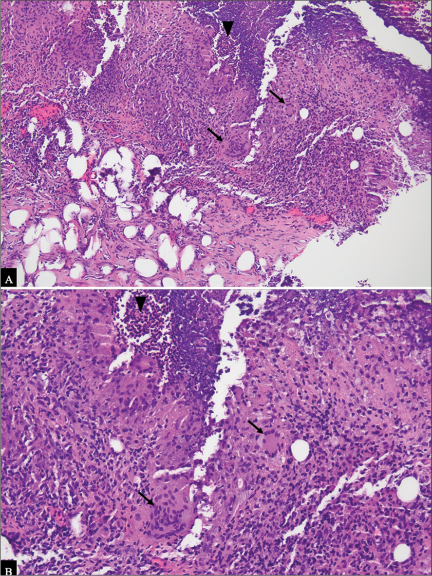
- (A) Routine hematoxylin and eosin stain (H&E) section of the lesion at 100× magnification demonstrates a mixed inflammatory lesion, comprised acute black inflammation (black arrowhead) and chronic inflammation with multinucleated giant cells (black arrows). (B) At 200×, the multinucleated giant cells (black arrows) and neutrophils (black arrowhead) are more easily visualized. Gram stain, and acid-fast bacillus stains were negative for bacteria, fungal elements, and acid-fast bacteria, respectively, within the tissue.
Interestingly, this patient’s skin infection following the ultrasound-guided percutaneous biopsy may have been a result of an infrequent but known complication. However, it has also been reported that GM often presents with multiple tender lumps forming micro abscesses at needle biopsy sites and may express sterile pus and resemble a skin infection,[5] similarly to this case in which the patient was treated for a suspected skin infection with bactrim. If antibiotic therapy fails and an infectious cause are ruled out, corticosteroids may be considered for the treatment. Corticosteroids, while effective in reducing inflammation, carry a large side effect risk, including immunosuppression and the potential for relapse once treatment is discontinued. Methotrexate has emerged as an effective alternative treatment for IGM, particularly in cases where corticosteroids are contraindicated or poorly tolerated. It has been used successfully as a steroid sparing agent, either alone or in combination with low-dose corticosteroids, in patients with recurrent or refractory IGM.[6] However, methotrexate therapy requires careful monitoring due to its potential side effects including hepatotoxicity, myelosuppression, and pulmonary toxicity. Surgery is reserved for refractive and aggressive cases. Recurrence may occur regardless of the treatment approach. Given this patient’s stability and mild symptoms, a conservative approach was deemed appropriate with a plan for close follow-up to monitor disease progression.
To the best of our knowledge, there has been only one other case published regarding IGM occurring secondary to breast biopsy, however associated with percutaneous ultrasound guided core biopsy in a 42-year-old patient with prolonged breast feeding,[5] but there have been other cases noting the development of IGM after other forms of trauma to the breast, such as post-ductoscopy.[7]
A more comprehensive review of the literature discusses how diagnosis of IGM remains a challenge, particularly in post-biopsy scenarios. It has been suggested that trauma, including from biopsy, may trigger an exaggerated immune response leading to granulomatous inflammation,[8] as seen in this case. While there are limited reports of IGM following percutaneous biopsy, this case adds to the growing body of evidence suggesting that such trauma can play a significant role in the development of IGM.
CONCLUSION
This case adds to the evolving understanding of IGM, particularly in the context of post-biopsy complications. Clinicians should remain vigilant in monitoring patients with a history of breast procedures, as trauma-induced GM may manifest years later. Continued research is warranted to elucidate the complex etiology of IGM and to optimize diagnostic and therapeutic strategies for this challenging condition.
Ethical approval
The Institutional Review Board approval is not required.
Declaration of patient consent
The authors certify that they have obtained all appropriate patient consent.
Conflicts of interest
There are no conflicts of interest.
Use of artificial intelligence (AI)-assisted technology for manuscript preparation
The authors confirm that there was no use of artificial intelligence (AI)-assisted technology for assisting in the writing or editing of the manuscript and no images were manipulated using AI.
Financial support and sponsorship
Nil.
References
- Idiopathic granulomatous mastitis: A case report and literature review. Clin Case Rep. 2023;11:e7819.
- [CrossRef] [Google Scholar]
- Predisposing factors for granulomatous lobular mastitis: A case-control study. Int J Womens Health. 2023;15:1063-75.
- [CrossRef] [Google Scholar]
- Idiopathic granulomatous mastitis: A rare confrontation. Cureus. 2021;13:e19420.
- [CrossRef] [Google Scholar]
- Overview of clinical aspects of breast disease In: Breast pathology (2nd ed). Netherlands: Elsevier; 2011. p. :28-33.
- [CrossRef] [Google Scholar]
- Idiopathic granulomatous mastitis masquerading as a postbiopsy abscess. Radiol Case Rep. 2015;8:773.
- [CrossRef] [Google Scholar]
- Effectiveness of methotrexate in idiopathic granulomatous mastitis treatment. Am J Med Sci. 2020;360:560-5.
- [CrossRef] [Google Scholar]
- Idiopathic granulomatous mastitis after ductoscopy: A case report. Int J Surg Case Rep. 2021;88:106540.
- [CrossRef] [Google Scholar]
- Idiopathic granulomatous mastitis and its mimics on magnetic resonance imaging: A pictorial review of cases from India. J Clin Imaging Sci. 2020;10:53.
- [CrossRef] [Google Scholar]


