
Translate this page into:
Giant Choledochal Cyst Mimicking Massive Gallbladder Hydrops in an Adult Patient: Multi Detector Computed Tomography and Magnetic Resonance Imaging Findings Correlated to Gross and Histopathological Findings
Address for correspondence: Dr. Joon-Il Choi, Department of Radiology, College of Medicine, Seoul St. Mary's Hospital, The Catholic University of Korea, 222 Banpo-daero, Seocho-gu, 137-701, Seoul, Republic of Korea. E-mail: dumkycji@gmail.com
-
Received: ,
Accepted: ,
This is an open-access article distributed under the terms of the Creative Commons Attribution License, which permits unrestricted use, distribution, and reproduction in any medium, provided the original author and source are credited.
This article was originally published by Medknow Publications & Media Pvt Ltd and was migrated to Scientific Scholar after the change of Publisher.
Abstract
Choledochal cysts are uncommon congenital anomalies of the biliary tree, commonly presenting in infancy, generally in the 1st year of life. Presentation in adult life is less common, accounting for 20% of cases. A 19-year-old female patient presented to the Emergency Department with severe abdominal distension, a palpable abdominal mass, mild jaundice and low grade fever. Ultrasound, computed tomography (CT) and magnetic resonance imaging of the abdomen showed a massive septated cystic lesion filling the entire abdomen with a significant mass effect on surrounding structures. Origin of the lesion was unclear and diagnosis included a giant mesenteric or duplication cyst, massive gallbladder with hydrops, biliary cystadenoma and giant choledochal cyst, among others. Final diagnosis was a Type IA choledochal cyst with massive asymmetric cystic dilatation of the extra-hepatic segments of the left hepatic duct with asymmetric dilatation of the right hepatic duct. Patient had an uneventful recovery after resection of the entire extrahepatic cyst and Roux-en-Y hepaticojejunostomy at the level of the hilum. In this article, we correlate CT and MRI findings to gross and histopathological findings of this giant Todani's Type IA choledochal cyst.
Keywords
Choledochal cysts
computed tomography
gallbladder hydrops
magnetic resonance cholangiopancreaticography

INTRODUCTION

Choledochal cysts are rare, congenital malformations of the biliary tree. They can be defined as aneurysmal dilatation of intra- and extra-hepatic ducts and are classified according to the involved segment of the biliary tree. The widely accepted classification system of choledochal cysts is proposed by Todani et al.[1] The most widely accepted etiology is anomalous pancreaticobiliary duct union, even though controversial.[2] Majority of cases are diagnosed in children. These lesions are one of the common cystic mass in the pediatric abdomen. However, only about 20% of the patients are adults.[3] The incidence of choledochal cysts is increasing due to the improvements in imaging techniques and choledochal cysts of adults get clinical attention due to various complications such as cholangitis, bile stasis, and malignancies of the biliary tree.[2]
At times, choledochal cysts may grow very large and differentiation from other cystic masses of the abdomen can be challenging. The correct diagnosis of choledochal cysts in pre-operative patients is essential since reconstruction of the biliary trees is mandatory in these patients. Here, we discuss the imaging features and correlate them to gross specimen in a young female with Type 1A giant choledochal cyst, which manifested as a huge cystic mass in the abdomen of unclear etiology.
RADIOLOGIC FEATURES
A previously healthy, 19-year-old female patient presented to the emergency room with intermittent abdominal pain and jaundice. Liver function test revealed an obstructive pattern of jaundice. Ultrasound (US) images showed a large septated cystic and tubular structure in the abdomen [Figure 1a]. Computed tomography (CT) scan was performed and it revealed a large, convoluted tubular and cystic structure in the right upper quadrant with associated dilated intrahepatic ducts [Figures 1b–d]. The long diameter of the cystic structure was over 23 cm with associated dilatation of intrahepatic ducts. There was no evidence of pancreatic ductal dilatation. No solid, enhancing components were noted in the wall of this cystic mass. Because of the compressive effect by this mass, the pancreatic head was not clearly seen and relationship between this mass and pancreatic duct was unclear.
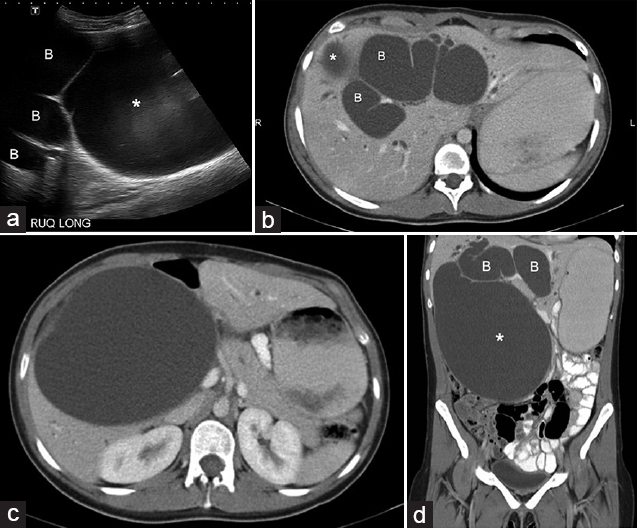
- 19-year-old female with intermittent abdominal pain and jaundice diagnosed with Type 1A giant choledochal cyst. (a) Ultrasound scan axial view shows a large cystic mass (*) and dilated bile ducts (B). The mass was very large and the relationship of the biliary tree to the mass was not clear on ultrasound. (b) Computed tomography (CT) scan, axial view of portal venous phase demonstrates dilated intrahepatic ducts (B). Superior portion of the cystic mass is visualized (*). (c) Computed tomography scan axial view at a lower level than 1b shows a large cystic mass (*) at the porta hepatis. (d) Coronal reformatted computed tomography image delineates a huge cystic mass in right upper quadrant (*). Dilated bile ducts are visualized (B).
Magnetic resonance cholangiopancreaticography (MRCP) demonstrated a huge septated cystic mass appearing to communicate with the intrahepatic ducts [Figure 2a]. Differential diagnoses include gallbladder hydrops, duplication cyst, mesenteric or peritoneal cyst, and a huge choledochal cyst.
In view of biliary dilatation [Figure 2b] a huge choledochal cyst was the most probable diagnosis.
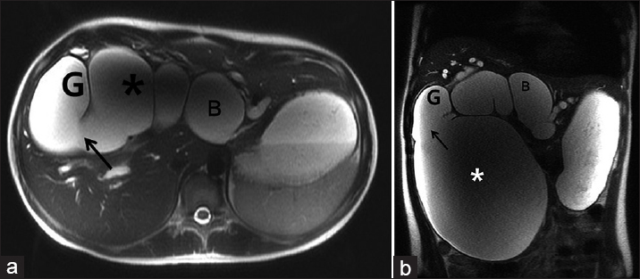
- 19-year-old female with intermittent abdominal pain and jaundice diagnosed with Type 1A giant choledochal cyst. a) Axial and b) coronal heavily T2-weighted images show a large cystic mass (*), gallbladder (G) and intrahepatic ducts (B). Communication of the gallbladder and cystic mass is visualized (arrow).
PATHOLOGIC FEATURES
Percutaneous transhepatic biliary drainage (PTBD) was performed. Roughly 2.5 L of bilious fluid was aspirated with subsequent decompression of the biliary tree.
Explorative laparotomy was scheduled and performed and severe dilatation of common bile duct and relatively small-sized gallbladder was found on surgery. Excision of the cystic mass and reconstruction surgery with hepaticojejunostomy were performed. Intraoperative finding was a massive, thick walled cystic lesion measuring over 20 cm in maximum dimension arising from the common bile duct. The cystic lesion involved entire extra-hepatic ducts and there was no stricture or soft-tissue masses. The gross specimen and histopathological examination confirmed the diagnosis of a giant choledochal cyst, Type IA according to Todani classification [Figure 3]. A schematic diagram shows the location of the cystic mass and its relationship to surrounding structures [Figure 4].
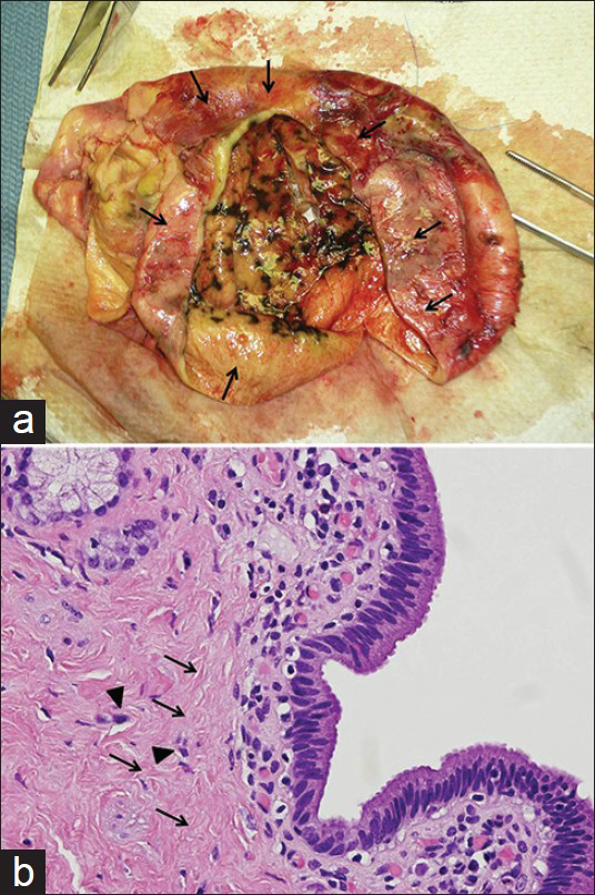
- 19-year-old female with intermittent abdominal pain and jaundice diagnosed with Type 1A giant choledochal cyst. a) The gross specimen obtained after excision of the cystic mass shows a massive, thick walled cystic lesion (arrows) measuring over 20 cm in maximum dimension arising from the left hepatic duct and confirms the diagnosis of a giant choledochal cyst. (b) H and E stained tissue (×400), shows the dilated bile duct with columnar epithelial lining, and dense fibrous wall; scattered smooth muscle and elastic fibers are present (arrows); mild infiltration of chronic inflammatory cells are also seen (arrowheads). There is no evidence of dysplasia or malignancy
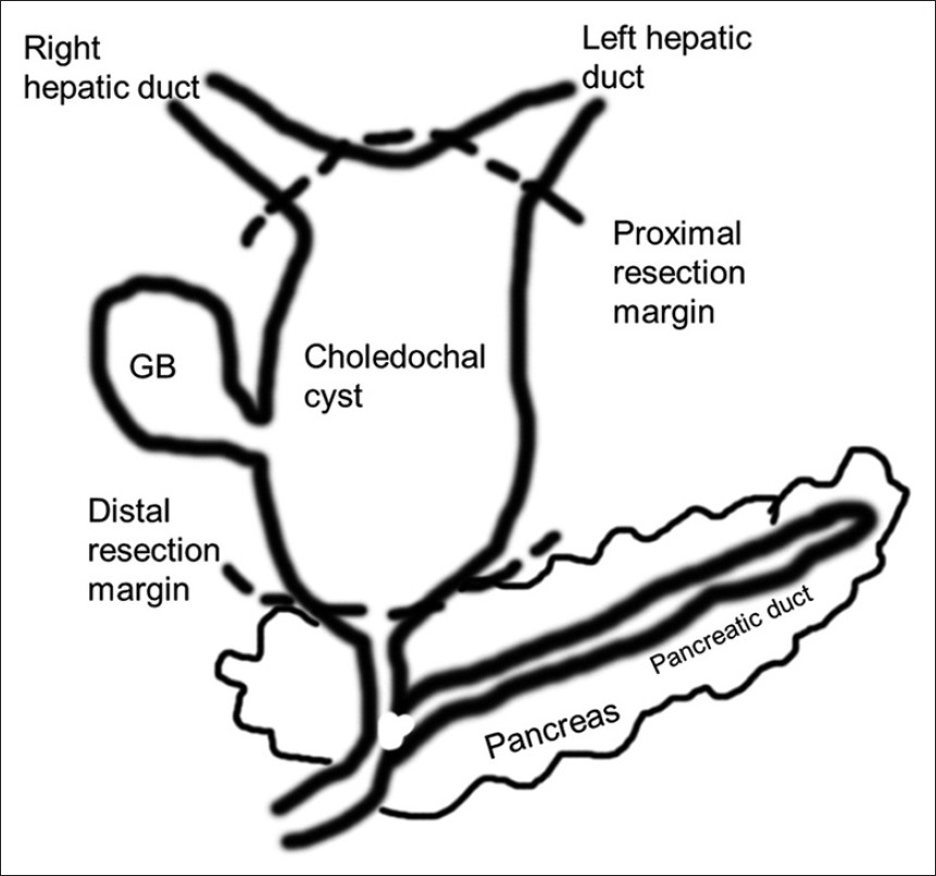
- Schematic diagram shows the location of the choledochal cyst and resection margin (dotted lines).
DISCUSSION
Choledochal cysts are very rare lesions seen in people in western countries. In Asian population the reported incidence rate is 1/1,000.[4] The incidence is increasing due to the use of the state-of-the-art imaging techniques such as multidetector CT (MDCT) or MRCP. Choledochal cysts in adults are rare and higher female predominance is reported.[5] Patients with choledochal cysts can have several complications and are considered as a high risk group for cholangiocarcinomas and surgical excision of the entire cyst is recommended.[6]
US is the primary imaging tool for evaluation, however, this modality has its limitations. Evaluating the entire biliary tree is difficult due to limited field of view and poor overview when compared with cross-sectional imaging modalities. Therefore, for accurate diagnosis, cross-sectional imaging modalities with a larger field of view are superior to US.
MDCT can obtain volumetric data sets and multiplanar reformations (MPR) are readily available. Furthermore, MRCP is the preferred imaging modality for the correct diagnosis of biliary anomalies.[7] MRCP is non-invasive and can be obtained without the use of contrast agents. 2D or 3D MRCP images clearly delineate biliary anomalies.
In adults patients, differential diagnosis for cystic masses at porta hepatis other than choledochal cysts include hepatic cysts, biliary cystadenomas, duplication cysts of the duodenum and colon, mesenchymal and peritoneal cysts and pseudocysts or cystic tumors from the pancreas. Presence of bile duct dilatation can be helpful for correct diagnosis. However, huge masses can compress adjacent structures and therefore intrahepatic ducts can be dilated simply due to mass effect. In those cases, MRCP can also be helpful to make a correct diagnosis because MRCP can delineate the relationship between cystic masses and adjacent structures.
The most common type of choledochal cyst is Type I and accounts for more than 75% of all choledochal cysts.[8] Type I cysts involve the extrahepatic bile ducts and can be divided into three subtypes; IA (cystic), IB (segmental) and IC (fusiform). Type I cyst should be differentiated from type IVA cysts which involve both intra- and extra-hepatic ducts. Type IVA cysts have strictures at the hepatic hilum. In our case, there was no stricture between intra- and extra-hepatic ducts and intrahepatic duct dilatation was continuous to the dilated extra-hepatic duct. Furthermore, after percutaneous transhepatic biliary drainage (PTBD) insertion, intrahepatic ducts are seen collapsed and the diagnosis of Type IA choledochal cyst can be made.
The causes of choledochal cysts remain unclear. One of the strongly suggested etiologies is reflux of pancreatic juice into the common bile duct and anomalous pancreaticobiliary ductal union (APBDU) can facilitate this phenomenon. Actually, strong association of presence of APBDU and choledochal cysts was reported by many authors. However, a case report of choledochal cyst in a 15-week-old fetus may refute this theory; i.e., 15-week-old fetus does not have pancreas tissue with exocrine function.[9] However, high incidence of choledochal cysts in patients with APBDU cannot be ignored.
Though endoscopic retrograde cholangiopancreaticography was considered as the gold standard for diagnosing abnormalities of the biliary tree and pancreatic ducts, MRCP is also excellent, non-invasive imaging modality for delineating and diagnosing congenital anomalies of pancreatic ducts and biliary trees including APBDU.[10] However, in our case, APBDU was not clearly demonstrated on MRCP because the choledochal cyst was too large and pancreas head was markedly compressed. Direct cholangiography via PTBD was not helpful also because the cystic mass was too large and filling of the pancreatic duct with contrast material was nearly impossible.
The ability to delineate APBDU is the strength of MRCP over MDCT. However, MDCT is cheap and convenient way of obtaining MPR images. Therefore, MDCT might be the primary imaging modality for huge cystic mass in the abdomen and MRCP can be a problem-solving tool.
As mentioned above, adult patients with choledochal cysts have a higher risk of cholangiocarcinomas and other complications are commonly encountered in these patients. Therefore, entire excision of choledochal cysts is recommended treatment option in most cases.
In our case, excision of the entire cyst and hepaticojejunostomy was performed. MRCP or MDCT also can be helpful for the planning of surgical excision and essential to pre-operative evaluation.
CONCLUSION
We report a giant, Type IA choledochal cyst presenting with a massive septated cystic mass in the abdomen in a young adult patient. State of the art cross-sectional imaging with MDCT or MRCP can help to reach the correct diagnosis and formulate a robust surgical plan.
Available FREE in open access from: http://www.clinicalimagingscience.org/text.asp?2013/3/1/45/120785
Source of Support: Nil
Conflict of Interest: None declared.
REFERENCES
- Congenital bile duct cysts: Classification, operative procedures, and review of thirty-seven cases including cancer arising from choledochal cyst. Am J Surg. 1977;134:263-9.
- [Google Scholar]
- Nationwide survey of cases of choledochal cyst. Analysis of coexistent anomalies, complications and surgical treatment in 645 cases. Surg Gastroenterol. 1984;3:69-73.
- [Google Scholar]
- Congenital choledochal cyst. Analysis of 1,433 patients in the Japanese literature. Am J Surg. 1980;140:653-7.
- [Google Scholar]
- Choledochal cysts: Differences between pediatric and adult patients. J Gastrointest Surg. 2010;14:1105-10.
- [Google Scholar]
- Congenital choledochal cyst. Analysis of 1,433 patients in the Japanese literature. Am J Surg. 1980;140:653-7.
- [Google Scholar]
- Antenatal diagnosis of choledochal cyst at 15 weeks’ gestation: Etiologic implications and management. J Pediatr Surg. 1989;24:936-8.
- [Google Scholar]
- Anomalies and anatomic variants of the biliary tree revealed by MR cholangiopancreatography. AJR Am J Roentgenol. 1999;173:1251-4.
- [Google Scholar]


