More results...

Generic selectors
Exact matches only
Search in title
Search in content
Post Type Selectors
Filter by Categories
Abdominal Radiology, Original Research
Abdominal Radiology, Pictorial Essay
Anthropology and Dental Radiology, Original Research
Breast Imaging, Case Report
Breast Imaging, Original Research
Breast Imaging, Pictorial Essay
Breast Imaging, Review Article
Cardiopulmonary Imaging, Case Report
Cardiopulmonary Imaging, Case Series
Cardiopulmonary Imaging, Education
Cardiopulmonary Imaging, Original Research
Cardiopulmonary Imaging, Pictorial Essay
Cardiopulmonary Imaging, Review Article
Case Report
Case Report, Breast Imaging
Case Report, Cardiopulmonary Imaging
Case Report, Diagnostic Radiology
Case Report, Gastrointestinal Imaging
Case Report, Genitourinary and Gynecologic Imaging
Case Report, Neuroradiology/Head and Neck Imaging
Case Report, Vascular and Interventional Radiology
Case Series
Case Series, Abdominal Radiology
Case Series, Diagnostic Radiology
Case Series, Musculoskeletal Imaging
Case Series, Vascular and Interventional Radiology
DENTAL ARTICLE
Dental Radiology, Case Report
Dental Radiology, Case Series
Dental Radiology, Original Research
Diagnostic Radiology, Case Report
Diagnostic Radiology, Case Series
Diagnostic Radiology, Original Research
Diagnostic Radiology, Pictorial Essay
Diagnostic Radiology, Review Article
Editorial
Education
Education, Diagnostic Radiology
Education, Imaging science
Education, Original Research
Emergency, Original Research
Erratum
Gastrointestinal Imaging, Case Report
Gastrointestinal Imaging, Case Series
Gastrointestinal Imaging, Original Research
Gastrointestinal Imaging, Pictorial Essay
General and Emergency Radiology, Original Research
General and Emergency Radiology, Review Article
Genitourinary and Gynecologic Imaging, Case Report
Genitourinary and Gynecologic Imaging, Original Research
Imaging Science, Original Research
Interventional Radiology, Original Research
Letter to Editor
Musculoskeletal Imaging, Case Report
Musculoskeletal Imaging, Original Article
Musculoskeletal Imaging, Original Research
Musculoskeletal Imaging, Pictorial Essay
Musculoskeletal Imaging, Review Article
Neuroradiology , Review Article
Neuroradiology Head and Neck Imaging, Pictorial Essay
Neuroradiology, Case Report
Neuroradiology, Pictorial Essay
Neuroradiology/Head and Neck Imaging, Case Report
Neuroradiology/Head and Neck Imaging, Case Series
Neuroradiology/Head and Neck Imaging, Original Research
Neuroradiology/Head and Neck Imaging, Review Article
Neuroradiology/Spine Imaging, Original Research
Notice of Retraction
Nuclear Medicine, Case Report
Nuclear Medicine, Original Research
Nuclear Medicine, Pictorial Essay
Original Article
Original Article, Neuroradiology
Original Research
Original Research Article
Original Research, Cardiopulmonary Imaging
Original Research, Dental Radiology
Original Research, Descriptive Study
Original Research, Diagnostic Radiology
Original Research, Gastrointestinal Imaging
Original Research, Genitourinary and Gynecologic Imaging
Original Research, Musculoskeletal Imaging
Original Research, Neuroradiology/Head and Neck Imaging
Original Research, Nuclear Medicine
Original Research, Vascular and Interventional Radiology
Pediatric Imaging, Case Report
Pediatric Imaging, Pictorial Essay
PICTORIAL ESSAY
Pictorial essay, Musculoskeletal Imaging
Pictorial essay, Neuroradiology/Head and Neck Imaging
PICTORIAL REVIEW
Radiologic-Pathologic Correlation
RADIOLOGICAL-PATHOLOGICAL CORRELATION
Radiology Business, Original Research
Research Article
Review Article
Review Article, Diagnostic Radiology
Review Article, Gastrointestinal imaging
Review Article, Musculoskeletal
Review Article, Musculoskeletal Imaging
Review Article, Vascular and Interventional Radiology
Technical Innovation
Technical Innovation, Cardiopulmonary Imaging
Technical Innovation, Gastrointestinal Imaging
Ultrasound, Case Report
Ultrasound, Original Research
Ultrasound, Review Article
Vascular and International Radiology, Case Report
Vascular and Interventional Radiology, Case Report
Vascular and Interventional Radiology, Case Series
Vascular and Interventional Radiology, Original Research
Vascular and Interventional Radiology, Pictorial Essay
Vascular and Interventional Radiology, Review Article
Vascular and Interventional Radiology, Short Communication
Vascular and Interventional, Original Research
View/Download PDF

Translate this page into:

CASE REPORT
2015
:5;
53
doi:
10.4103/2156-7514.166355

Ganglioglioma of the Spinal Cord

Department of Imaging Sciences, University of Rochester Medical Center, Rochester, New York, USA
Department of Pathology and Laboratory Medicine, University of Rochester Medical Center, Rochester, New York, USA
Department of Pathology and Laboratory Medicine, Children's Hospital Los Angeles, Los Angeles, California, USA

Address for correspondence: Dr. Daniel C. Oppenheimer, Department of Imaging Sciences, University of Rochester Medical Center, 601 Elmwood Avenue, Rochester, NY 14642, USA. E-mail: Daniel_Oppenheimer@URMC.Rochester.edu

Licence

This is an open access article distributed under the terms of the Creative Commons Attribution-NonCommercial-ShareAlike 3.0 License, which allows others to remix, tweak, and build upon the work non-commercially, as long as the author is credited and the new creations are licensed under the identical terms.

Disclaimer:
This article was originally published by Medknow Publications & Media Pvt Ltd and was migrated to Scientific Scholar after the change of Publisher.

Abstract

Ganglioglioma is a rare tumor consisting of neoplastic glial and neuronal elements. It accounts for only 0.5% of all primary central nervous system (CNS) neoplasms. We report an unusual case of extensive intramedullary thoracic spinal cord ganglioglioma in a 14-month-old girl who underwent subtotal resection followed by adjuvant chemotherapy. The epidemiology, histopathologic features, imaging findings, treatment, and prognosis are subsequently reviewed.

Keywords

Ganglioglioma
intramedullary
surgical debulking

INTRODUCTION

Ganglioglioma is a rare, slow-growing primary central nervous system (CNS) tumor which most frequently occurs in the temporal lobes, typically resulting in seizures.[1] Ganglioglioma of the spinal cord is exceedingly rare – only approximately 70 cases of spinal cord ganglioglioma have been reported, mostly in children or young adults.[2] The gold Standard of treatment is gross total resection; however, the optimal treatment in the setting of subtotal resection has not been clearly established.

CASE REPORT

A 14-month-old female child was initially evaluated for abnormal neck posture and difficulty turning her head to the right and upward. The patient was noted to perform compensatory maneuvers such as using her arms to push herself up to look around rather than simply lifting her head. Similarly, the patient was observed to make a 270° turn to the left rather than making a simple 90° turn to the right. Physical exam was notable for leftward deviation of the head with increased bulk and tone in the right sternocleidomastoid muscle with associated right shoulder elevation. Dextroconvex thoracic scoliosis was also present. Attempts at moving the neck provoked the patient to cry. She had good fine motor coordination for her age, but was not yet walking unassisted. No additional motor, sensory, gait, or reflex abnormalities were present. The physical exam finding of scoliosis prompted referral to orthopedic surgery and an MRI was subsequently obtained.

The MR revealed a long segment intramedullary lesion extending from T1 to T11 levels with corresponding cord expansion, heterogeneous enhancement following gadolinium administration [Figure 1], isointense T1 signal, and heterogeneously hyperintense T2 signal [Figure 2]. There was associated syringomyelia at the rostral and caudal aspects of the enhancing tumor. Additionally, there was T2 hyperintensity in the medulla and upper cervical spine as well as in the caudal spinal cord without associated enhancement.
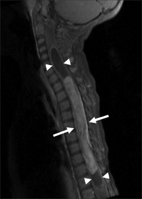
14-month-old female child with spinal cord ganglioglioma. Sagittal T1 fat-suppressed gadolinium contrast-enhanced MR image of the cervical and thoracic spine shows a long segment enhancing intramedullary lesion (arrows) extending from T1 to T11 with cord expansion and syringomyelia (arrowheads) at the rostral and caudal aspects of the enhancing tumor.
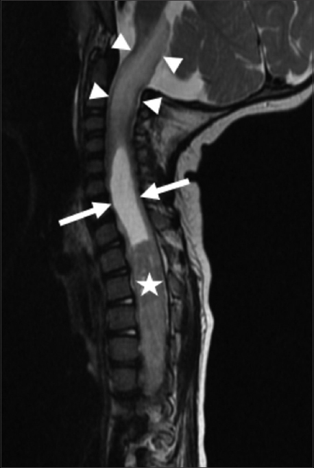
Figure 1
14-month-old female child with spinal cord ganglioglioma. Sagittal T1 fat-suppressed gadolinium contrast-enhanced MR image of the cervical and thoracic spine shows a long segment enhancing intramedullary lesion (arrows) extending from T1 to T11 with cord expansion and syringomyelia (arrowheads) at the rostral and caudal aspects of the enhancing tumor.
14-month-old female child with spinal cord ganglioglioma. Sagittal T2-weighted MR image of the cervical and thoracic spine shows syringomyelia (arrows) rostral to the intramedullary tumor (star). Abnormal hyperintense T2 signal and expansion is noted in the upper cervical spinal cord and brainstem (arrowheads).
Figure 2
14-month-old female child with spinal cord ganglioglioma. Sagittal T2-weighted MR image of the cervical and thoracic spine shows syringomyelia (arrows) rostral to the intramedullary tumor (star). Abnormal hyperintense T2 signal and expansion is noted in the upper cervical spinal cord and brainstem (arrowheads).

The patient subsequently underwent thoracic en bloc T1–T12 laminectomy and debulking of thoracic intramedullary spinal cord tumor with use of electromyography, somatosensory evoked potentials, motor evoked potentials, and D-wave neuromonitoring. Tumor was debulked using a combination of posterior myelotomy and Cavitron Ultrasonic Surgical Aspirator (CUSA). Unfortunately, during exploration, it was determined that the tumor was variably interspersed with normal spinal cord. Therefore, only subtotal resection could be safely achieved with an intraoperative estimation of approximately 60–70% of the tumor resected.

Postoperatively, the patient maintained normal function of all extremities and MR showed reduced size of the enhancing tumor, less cord expansion, and improved T2 hyperintensity in the adjacent cord. However, due to the significant residual tumor burden, chemotherapy was initiated with Carboplatin and Vincristine. Unfortunately, follow-up imaging 3 months later showed increased enhancing intramedullary tumor with worsening cord expansion and T2 hyperintensity. Chemotherapy was changed to Vinblastine. Despite imaging evidence of progression of disease, the patient displayed no new neurologic deficits, although scoliosis and abnormal neck posture persisted. Additional follow-up MR 6 months after initiating Vinblastine showed improved cord edema and less-enhancing tumor. At the time of this writing, the patient is able to walk and jump unassisted, has started talking, and continues to meet appropriate developmental milestones.

Sections from the surgical excision revealed diffuse glial fibrillary acidic protein (GFAP)-immunoreactive glioma with collections of atypical, variably binucleate synaptophysin-immunoreactive cells with no Neu-N or neurofilament immunoreactivity [Figures 35]. There was focal necrosis and calcification, but no eosinophilic granular bodies. There was no immunoreactivity to isocitrate dehydrogenase gene 1 (R132) or p53. Cluster of differentiation 34 (CD34) immunostaining was confined to the blood vessels. The reticulin showed no notable pericellular staining. There were rare mitoses, but the Ki-67 labeling was variable and focally 15%. Polymerase chain reaction revealed no BRAF v600E mutation, NMYC or epidermal growth factor receptor gene amplification, and fluorescence in situ hybridization found no chromosome 10q loss. A diagnosis of glioneuronal tumor consistent with WHO grade I ganglioglioma was made. This diagnosis was confirmed with a second opinion obtained from a different academic institution.
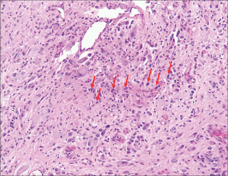
14-month-old female child with spinal cord ganglioglioma. Hematoxylin and eosin stained (×200).surgical resection specimen shows the tumor consisting of numerous dysplastic and binucleate ganglion cells in clusters (arrows).
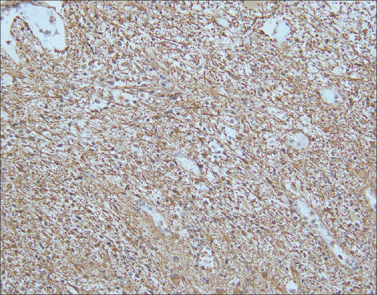
Figure 3
14-month-old female child with spinal cord ganglioglioma. Hematoxylin and eosin stained (×200).surgical resection specimen shows the tumor consisting of numerous dysplastic and binucleate ganglion cells in clusters (arrows).
14-month-old female child with spinal cord ganglioglioma. Immunohistochemistry performed on the surgical resection specimen shows fibrillary GFAP-immunoreactive astrocytes (brown) spanning the tumor stroma (anti-GFAP immunohistochemical staining done with diaminobenzidine chromagen and hematoxylin counterstain; original magnification ×200).
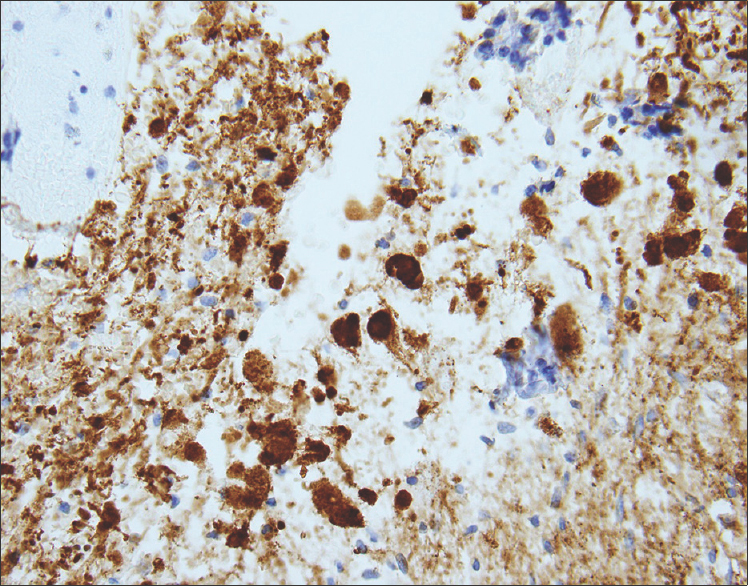
Figure 4
14-month-old female child with spinal cord ganglioglioma. Immunohistochemistry performed on the surgical resection specimen shows fibrillary GFAP-immunoreactive astrocytes (brown) spanning the tumor stroma (anti-GFAP immunohistochemical staining done with diaminobenzidine chromagen and hematoxylin counterstain; original magnification ×200).
14-month-old female child with spinal cord ganglioglioma. Immunohistochemistry performed on the surgical resection specimen shows dysplastic and binucleate ganglion cells with extensive synaptophysin immunoreactivity in somas and processes (brown) (anti-synaptophysin immunohistochemical staining done with diaminobenzidine chromagen and hematoxylin counterstain; original magnification ×200).
Figure 5
14-month-old female child with spinal cord ganglioglioma. Immunohistochemistry performed on the surgical resection specimen shows dysplastic and binucleate ganglion cells with extensive synaptophysin immunoreactivity in somas and processes (brown) (anti-synaptophysin immunohistochemical staining done with diaminobenzidine chromagen and hematoxylin counterstain; original magnification ×200).

DISCUSSION

Ganglioglioma is a rare CNS tumor consisting of neoplastic glial and neuronal elements.[1] The temporal lobe is the most frequent site of involvement; brainstem and spinal cord gangliogliomas are much less common. Within the spinal cord, ganglioglioma most frequently involves the cervical cord, although cases have been reported involving the thoracic and lumbar spine and the entire length of the spinal cord.[3] Spinal cord ganglioglioma has been observed in a wide age range of patients from newborn to elderly, but most cases occur in children or young adults. No gender, race, or ethnic association has been identified. Patients typically present with progressive myelopathy in addition to other neurological deficits related to location of the lesion. Motor and sensory defects, gait abnormalities, bowel and bladder dysfunction, and pain have all been described in patients with spinal ganglioglioma.[4]

Gangliogliomas are generally slow growing, and therefore, tumors are often large and detected at a late stage due to insidious progression. Only one case of spinal cord ganglioglioma presenting with acute neurological deficit has been described, which was attributed to tumor hemorrhage. In our case, the patient had several months of symptoms including scoliosis and neck deviation.

CT is generally not a recommended modality for diagnosis and evaluation of spinal cord tumors, particularly in the pediatric population. Evaluation with MR most commonly demonstrates a circumscribed solid or mixed solid and cystic mass spanning a long segment of the cord with hypointense T1 signal and hyperintense T2 signal in the solid component. Enhancement patterns are highly variable, ranging from minimal to marked, and may be solid, rim, or nodular. Adjacent cord edema and syringomyelia and peritumoral cysts may be present in addition to reactive scoliosis.[5] Our case demonstrated many typical imaging findings including long segment solid enhancing tumor, T2 hyperintensity, scoliosis, and syrinx formation.

It is nearly impossible to differentiate ganglioglioma from other more common intramedullary neoplasms based on imaging alone. Astrocytoma and ependymoma are more familiar intramedullary tumors which share many similar features to ganglioglioma, including T2 hyperintensity, enhancement, tumoral cysts, and cord edema. Poorly defined margins may be more suggestive of astrocytoma, while a central location in the spinal cord, hemorrhage, and hemosiderin staining are often seen with ependymoma. Hemangioblastoma and paraganglioma are less usual intramedullary tumors, but since they are more frequently encountered than ganglioglioma, they should also be included in the differential diagnosis.

Histologically, ganglioglioma is composed of both neoplastic glial and ganglion cells which are disorganized, variably cellular, and non-infiltrative. Occasionally, it may be challenging to differentiate ganglion cell tumors from an infiltrating glioma with entrapped neurons. The presence of neoplastic ganglion cells forming abnormal clusters, the presence of binucleation and dysmorphic neurons are helpful clues favoring ganglioglioma over glioma. The glial component of ganglioglioma includes fibrillary astrocytes with varying degrees of cellular atypia. The neoplastic neuronal components are often clustered or irregularly oriented. Fibrovascular stroma confined to the neuronal component, perivascular lymphocytic infiltrates, and small foci of calcification are common, as is immunopositivity for synaptophysin, neuron-specific enolase, and chromogranin A. Elevated Ki-67 and p53 labeling index is associated with more aggressive tumor behavior in both children and adults with gangliogliomas. The rare occurrence of malignant transformation is confined to the glial cell population, and is characterized by increased cellularity and mitotic activity, endothelial proliferation, and necrosis.[6]

Gangliogliomas are generally benign WHO grade I tumors; the presence of anaplastic changes in the glial component is considered to represent WHO grade III (anaplastic ganglioglioma). Criteria for WHO grade II have been suggested, but are not established.[7] Malignant transformation of spinal ganglioglioma has been seen in only a select few cases.[3] Poor prognostic factors for adults with gangliogliomas include older age at diagnosis, male sex, and malignant histologic features.

Definitive treatment for ganglioglioma requires gross total surgical resection, and a good prognosis is generally expected when this is achieved.[8] However, indistinct tumor margins and the desire to preserve normal spinal cord tissue, motor and sensory function may preclude complete resection of tumor. According to a series by Lang et al., reviewing several patients with resected spinal cord ganglioglioma, the 5- and 10-year survival rates after total resection were 89% and 83%, respectively.[4] In that study, patients with spinal cord ganglioglioma had a 3.5-fold higher relative risk of tumor recurrence compared to patients with supratentorial ganglioglioma. It has been recognized that postoperative results correlate closely with preoperative neurological status as well as the ability to achieve complete resection.[9] With the exception of WHO grade III anaplastic ganglioglioma, radiation therapy is generally regarded to have no role in the treatment of ganglioglioma.[10] In fact, radiation therapy may induce malignant transformation of a recurrent ganglioglioma several years later. Adjuvant chemotherapy is also typically reserved for anaplastic ganglioglioma, but has been used anecdotally in partially resected low grade spinal cord gangliogliomas which show evidence of disease progression, such as in the case presented above.

CONCLUSION

We present a case of extensive thoracic spinal ganglioglioma in a 14-month-old girl, whose clinical symptoms remained stable after subtotal resection and adjuvant chemotherapy, despite imaging evidence of tumor growth. While ganglioglioma is a very rare tumor of the spinal cord, it must be considered in the differential diagnosis of an intramedullary tumor. Early recognition, diagnosis, and treatment is imperative to improve prognosis and minimize neurological sequelae. Unfortunately, the diagnosis is often delayed in clinical practice due to the slow growth and indolent nature of this tumor. The benefit of radical resection must be weighed against the risk of further worsening neurological function, particularly in young patients who have a longer life expectancy. Further research on the optimal treatment of partially resected and recurrent spinal cord ganglioglioma is necessary.

Financial support and sponsorship

Nil.

Conflicts of interest

There are no conflicts of interest.

REFERENCES

  1. , , , , , , . Childhood ganglioglioma and medically intractable epilepsy.A clinicopathological study of 15 patients and a review of the literature. Pediatr Neurosurg. 1995;22:181-8.
    [Google Scholar]
  2. , , , . Ganglioglioma of the spinal cord: Report of two rare cases and review of the literature. Neurosurgery. 1997;41:1410-6.
    [Google Scholar]
  3. , , . Ganglioglioma of the entire spinal cord. Childs Brain. 1980;6:274-80.
    [Google Scholar]
  4. , , , , , , . Central nervous system gangliogliomas. Part 2: Clinical outcome. J Neurosurg. 1993;79:867-73.
    [Google Scholar]
  5. , , , , , , . MR of spinal cord ganglioglioma. AJNR Am J Neuroradiol. 1998;19:879-87.
    [Google Scholar]
  6. , , , , , , . Ganglioglioima: An ultrastructural and immunohistochemical study. Cancer. 1997;79:989-1003.
    [Google Scholar]
  7. , , , , . WHO Classification of Tumours of the Central Nervous System. (4th ed). Lyon: International Agency for Research on Cancer; . p. :103.
    [Google Scholar]
  8. , , , , , , . Pediatric low-grade ganglioglioma: Epidemiology, treatments, and outcome analysis on 348 children from the surveillance, epidemiology, and end results database. Neurosurgery. 2015;76:313-20.
    [Google Scholar]
  9. , , , , , , . Clinical outcome of pediatric gangliogliomas: Ninety-nine cases over 20 years. Pediatr Neurosurg. 1997;27:203-7.
    [Google Scholar]
  10. , , , . Ganglioglioma of the spinal cord: Report of two cases and review of literature. J Clin Neurosci. 2004;11:199-203.
    [Google Scholar]
Show Sections