
Translate this page into:
Ewing's Sarcoma of the Finger
Address for correspondence: Dr. Veysel Kaplanoglu, Department of Radiology, Keçiören Education and Research Hospital, Ankara, Turkey. E-mail: hatice.altnkaynak@yahoo.com.tr
-
Received: ,
Accepted: ,
This is an open-access article distributed under the terms of the Creative Commons Attribution License, which permits unrestricted use, distribution, and reproduction in any medium, provided the original author and source are credited.
This article was originally published by Medknow Publications & Media Pvt Ltd and was migrated to Scientific Scholar after the change of Publisher.
Abstract
Ewing's sarcoma is a mesenchymal cell tumor usually seen in long bones but very rarely seen in the bones of a finger. Swelling and pain are the most common complaints of the affected finger. In radiological imaging, it may be seen as permeative bone destruction accompanied by a soft tissue component or an expansile bone lesion. A 27-year-old right-hand dominant female patient presented with a swelling on the proximal phalanx of her right 3rd finger that had existed for 3 years. However, the mass started to gradually increase in size and the pain worsened over a period of 5 weeks. The mass was excised under regional intravenous anesthesia and Ewing's sarcoma was confirmed following a histopathological evaluation. No local recurrence or metastasis was detected 1 year after surgery. Since Ewing's sarcoma is rarely seen in the finger, we present this case with its radiological and clinical findings.
Keywords
Ewing's sarcoma
magnetic resonance imaging
phalanx

INTRODUCTION

Ewing's sarcoma (ES) was first described in 1921.[1] It is the second most common primary bone tumor of childhood and adolescence.[2] It usually affects long bones; however, it is very rarely seen in the hand, especially in the fingers.[1] Compared to the typical radiological findings on other localizations, this sarcoma usually shows different radiological features when it appears on the hand or foot.[3] In this paper, we present this case of ES that was treated surgically.
CASE REPORT
A 27-year-old right-hand dominant female patient presented with a 3-year history of swelling on the proximal phalanx of her right 3rd finger. However, the mass started to gradually increase in size and the pain worsened over a period of 5 weeks. There was no recent history of trauma, fever, or weight loss. On physical examination, the mass was immobile and fixed. All the laboratory findings were within the normal range. Conventional radiographs revealed preserved bone contours, surrounding soft tissue swelling, and increase in the opacity of the proximal phalanx of the 3rd finger [Figure 1]. Magnetic resonance imaging (MRI) revealed a homogeneous soft tissue lesion about 4.7 × 3.3 cm in size on the right 3rd proximal phalanx, next to the flexor tendon. The lesion showed significant enhancement with focal lobulation on the proximal point. It was hypointense on T1-weighted (T1W) images and hyperintense on T2-weigthed (T2W) images [Figure 2a–c]. The neighboring bone cortex was natural in appearance and no metastasis was detected in the preoperative computed tomography (CT) of the thorax and in the full-body scintigraphy. The lesion was surgically excised and a microscopic evaluation showed tumor cells that were round in shape with vesicular nuclei, finely dispersed chromatin, narrow cytoplasm, and stratified monotonous cells with indistinct borders. In places, the cytoplasm could be clearly seen due to the glycogen content [Figure 3]. The immunohistochemical study showed widespread and powerful staining of the cells with cluster of differentiation 99 (CD99) antigen. Cytokeratin and epithelial membrane antigen (EMA) showed focal staining. The cells stained positive for vimentin, but did not stain positive for S-100, cytokeratin 7, cytokeratin 19, desmin, actin, CD34, factor 8, and calretinin. The Ki67 proliferation index was 30%. The pathological diagnosis was Ewing's sarcoma. The patient received postoperative chemotherapy consisting of VAC + IE [cyclophosphamide (C), actinomycin (A), vincristine (V), ifosfamide (I), and etoposide (E)]. On follow-up after 1 year, no postoperative local recurrence or metastasis was detected.
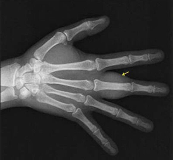
- 27-year-old right-hand dominant female patient with a swelling on the proximal phalanx of her right 3rd finger diagnosed as Ewing's sarcoma. X-ray of the right hand AP view shows swelling of the soft tissue and increase in the density of the proximal phalanx of the 3rd finger (arrow).
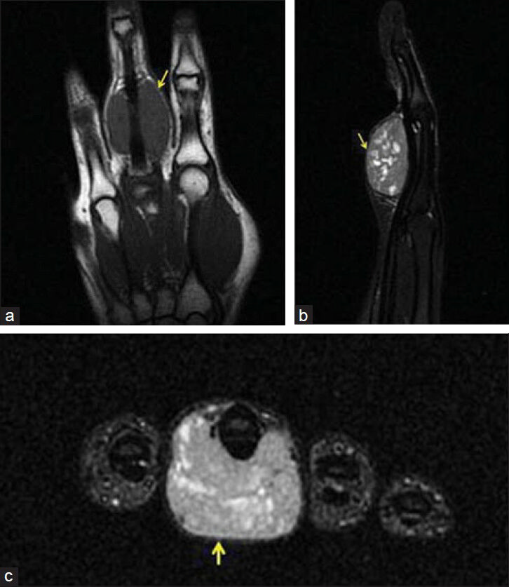
- 27-year-old right-hand dominant female patient with a swelling on the proximal phalanx of her right 3rd finger diagnosed as Ewing's sarcoma. (a) T1W coronal, (b) T2W sagittal, (c) T1W axial contrast enhanced images of the right hand 3rd finger, proximal phalanx palmar side, neighboring the flexor tendon, show a lesion hypointense on T1W (arrow), hyperintense on T2W (arrow), with contrast enhancement (solid arrow).
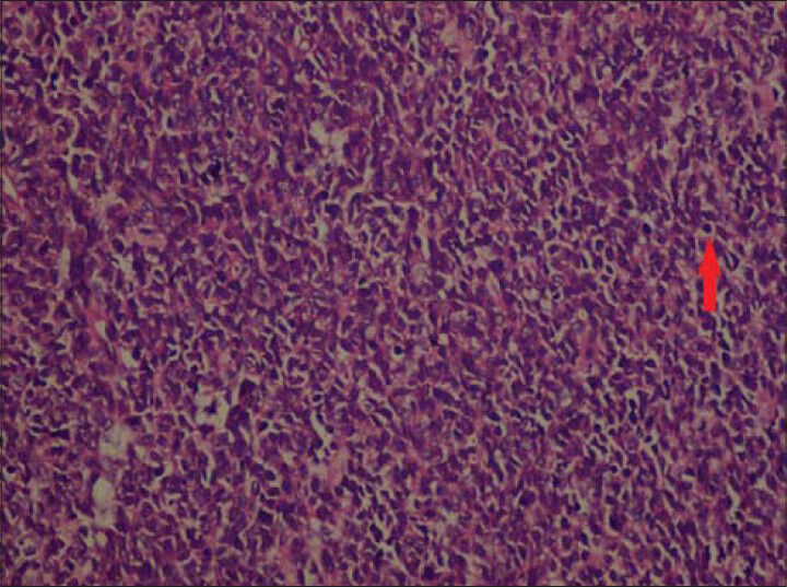
- 27-year-old right-hand dominant female patient with a swelling on the proximal phalanx of her right 3rd finger diagnosed as Ewing's sarcoma. Excised tissue stained with hematoxylin and eosin (×400) shows Ewing's sarcoma tumor cells in parts with clear appearance due to presence of glycogen (arrow).
DISCUSSION
Ewing's sarcoma is a mesenchymal cell tumor.[1] It is usually seen in the first or second decade of life, generally affecting long tubular bones and the pelvis.[2] It is very rarely seen in the bones of the hand. Less than 1% of the cases diagnosed with ES are localized in the hand and the wrist. The most common locations in the hand are the metacarpal and proximal phalanxes.[3] The thumb (28%) and the middle finger (28%) are most commonly affected.[1] Men (69%) are affected more than the women, with the average age of occurrence being 18.5 years (5 months–51 years).[1] Pain and swelling are the most common complaints of the affected finger.[2] At the beginning of the clinical presentation, the patient's general health condition is good with no fever or weight loss being present.[4] In the current case, the patient was in good health, with the only complaints being pain and swelling of the finger.
Detection of the site and size of a primary tumor is very important for the planning and application of optimal local treatment. Therefore, X-ray and MRI of the primary site, chest CT, and bone scintigraphy should be performed for staging. An X-ray will show typical destructive lesions with indistinct margins, onion-skin–type periosteal reactions and soft tissue masses in the diaphysis or, more rarely, in the metaphyseal/diaphyseal region.[5] A sclerotic appearance may be seen on smooth bones. MRI is an ideal method for evaluating soft tissues, the intramedullary involvement of the disease, and the involvement of the primary tumor in the surrounding soft tissues. Bone scintigraphy assists in detecting the bone involvement and, to an extent, the amount of intramedullary involvement. Conventional Tc99 bone scintigraphy is ideal for metastatic disease scanning.[6]
ES is clinically characterized by fever and an increase in the erythrocyte sedimentation rate. This may be confused with tuberculous dactylitis.[3] Radiologically, it may be seen as bone destruction and expansile lesion. On X-ray, it may be confused with osteomyelitis due to bone destruction.[1] Furthermore, tuberculous dactylitis is characterized by expansile lesion of the hand and absence of periosteal reaction. However, permeating bone destruction, onion-skin–type periosteal reaction, large soft tissue swelling, and cystic lesions causing thickening of the bone are only seen in ES.[7] Osteosarcoma is more commonly found in the metaphysis, while ES occurs in the diaphysis.[8] Furthermore, an increase of alkaline phosphatase (ALP) is seen in osteosarcoma, but not in ES. Differing from ES, in a primitive neuroectodermal tumor, the large soft tissue components reach inside the bone. Askin tumors, however, are only seen on the chest wall.[9] The tumor may be clinically confused with infection because it causes pain and local inflammatory symptoms. In addition, metastatic diseases and hematological malignancies should be considered in the differential diagnosis.[9]
Radiological features such as permeating bone destruction and laminated or spiculated periosteal reaction may not be seen in hand lesions.[8] Bone expansion, cystic or honeycomb appearance, periosteal reaction, and cortical thickening of the bone are less seen in the ES lesion on the hand or the foot.[8] Regardless of the treatment method, the localization of the tumor on the hand is an important prognostic factor. When the distal parts of the extremities are affected, the survival rate is much higher. The best treatment combination is radical excision and postoperative chemotherapy. This treatment approach is successful in tumors of the fingers. Radiotherapy is useful when radical surgical excision is not performed and in partial response to chemotherapy. Currently, the most effective treatment method is a combination of surgery and chemotherapy.[1]
CONCLUSION
Although fingers are a rare site for localization of Ewing's sarcoma, ES should be considered in the differential diagnosis of lesions in this location.
Available FREE in open access from: http://www.clinicalimagingscience.org/text.asp?2014/4/1/57/143420
Source of Support: Nil
Conflict of Interest: None declared.
REFERENCES
- Ewing's sarcoma of the finger: Report of two cases and literature review. Orthop Traumatol Surg Res. 2012;98:233-7.
- [Google Scholar]
- Ewing's sarcoma of the distal phalanx of the little finger. J Hand Surg Eur. 2008;33:81-2.
- [Google Scholar]
- Ewing sarcoma of thumb: Report of a case and review of the literature. Ann Chir Plast Esthet. 2004;49:378-82.
- [Google Scholar]
- Preoperative evaluation and monitoring chemotherapy in patients with high-grade osteogenic and Ewing's sarcoma: Review of current imaging modalities. Skeletal Radiol. 1998;27:57-71.
- [Google Scholar]
- Detection of malignant bone tumors: MR imaging vs scintigraphy. AJR Am J Roentgenol. 1990;155:1043-8.
- [Google Scholar]
- Ewings sarcoma of the small bones of hands and feet. Indian J Radiol Imaging. 2002;12:403-5.
- [Google Scholar]
- Classics in oncology. Diff use endothelioma of bone. James Ewing. Proceedings of the New York Pathological Society, 1921. CA Cancer J Clin. 1921;22:95-8.
- [Google Scholar]


