
Translate this page into:
Elastography-targeted Thyroid Nodule Aspiration: A Novel Approach
-
Received: ,
Accepted: ,
This is an open access article distributed under the terms of the Creative Commons Attribution-NonCommercial-ShareAlike 3.0 License, which allows others to remix, tweak, and build upon the work non-commercially, as long as the author is credited and the new creations are licensed under the identical terms.
This article was originally published by Medknow Publications & Media Pvt Ltd and was migrated to Scientific Scholar after the change of Publisher.
Abstract
Objectives:
Since 2009, the rate of nondiagnostic (ND) thyroid nodule fine-needle aspiration (FNA) has ranged from 2% to 20%. A ND result can cause further patient morbidity secondary to repeated procedures and delay in diagnosis. The use of real-time strain elastography (RTE) in determining nodule malignant risk has gained considerable focus recently. A less studied area where RTE may prove beneficial is its role in targeting areas for FNA. Our hypothesis is that FNA performed in concurrence with RTE will show a decreased rate of ND results leading to fewer repeated FNA.
Materials and Methods:
The Institutional Review Board approval was obtained. A retrospective review of all thyroid nodule FNA from January 1, 2011, to January 1, 2014, was performed with review of nodule size, presence of microcalcifications, vascularity, solid components, patient age, and gender. Cases were separated based if RTE was done before FNA or not. Pathology reports were reviewed to assess for specimen adequacy. Statistical comparison was performed using SAS analysis software.
Results:
A total of 221 specimens were reviewed, with RTE performed on 140 cases (63.4%). Both groups were similar in demographics and previously described nodule characteristics. The ND rate when RTE was not performed was 16% (13/68) compared to 10% when RTE was performed (14/126). The difference was not found to be statistically significant, P = 0.205.
Conclusions:
The presence of an elastogram failed to demonstrate a significant decrease in ND FNA rates although these results may be secondary to study design. Further evaluation with prospective trials using larger sample size may ultimately detect increased accuracy of RTE-targeted FNA.
Keywords
Elastography
fine-needle aspiration
nondiagnostic
thyroid nodule
ultrasound


Introduction
Ultrasound-guided fine-needle aspiration (FNA) has become the gold standard for differentiating benign and malignant thyroid nodules due to the suboptimal accuracy using gray scale features alone. Besides for mild associated pain, the procedure is usually safe and well tolerated. Development of postprocedural hematoma has been reported from 1.9% to 6.4%, with seven reported cases of life-threatening hematomas relating to airway obstruction.[1] The true rate of complications following thyroid nodule FNA is likely much higher though as most go undocumented and unpublished.
Although FNA plays an essential role in thyroid nodule evaluation, the potential for a nondiagnostic (ND) specimen can lead to repeat procedures, increasing patient morbidity, cost, chance for complication, and delay in diagnosis. Recently, thyroid nodule FNA pathological reporting has been standardized based on the Bethesda system for reporting thyroid cytopathology adopted by the American Society for Clinical Pathology (ASCP). Based on their criteria, an adequate specimen needs to contain at least six groups of follicular cells with each group composed of at least 10 cells each. An ND specimen can be secondary to obscuring blood, overlying thick smears, air drying of alcohol-fixed smears, or inadequate number of follicular cells. According to the ASCP, ND specimens occur in 2%–20% of cases but ideally should be limited to no more than 10% of all thyroid nodule FNAs.[2]
Recently, the use of ultrasound strain elastography (USE) has emerged as a promising diagnostic tool in differentiating benign and malignant thyroid. A novel approach aimed at decreasing the rate of ND nodule FNA maybe to use elastography as a map, targeting biopsy needles to areas of lower strain (i.e., stiffer). Several papers have been published illustrating increased detection of prostate cancer using USE-targeted interventions through a transrectal approach.[34]
Our study was performed to evaluate the role of USE-targeted FNA in decreasing the rates of ND aspirates. We performed a review of all thyroid nodule FNA performed over a 3-year period comparing FNA results when an elastogram was available before FNA or not. Our hypothesis is that the presence of an elastogram before FNA would decrease the rate of ND specimens.
Materials and Methods
This was a single-center retrospective review. Approval for this review was obtained from the hospital's Institutional Review Board. The study was Health Insurance Portability and Accountability compliant, and informed consent was waived.
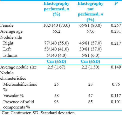
Between January 2011 and January 2014, a total of 225 patients underwent ultrasound-guided thyroid nodule FNA. Among them were 171 females and 54 males with ages ranging from 27 to 90. The demographical data recorded included patients' age and gender as well as which thyroid lobe contained the nodule biopsied [Table 1]. Gray scale nodule characteristics that were recorded included the presence of microcalcifications, vascularity, solid components, and nodule size [Table 1]. The decision to perform USE was based strictly on suspicious gray scale characteristics, and guidelines set forth by the Society of Radiologists in Ultrasound.[5]
During this period, USE for thyroid nodules was performed in cases where the available technician was comfortable and experienced with the elastography software. When elastography was performed, the applied transducer pressure was standardized by a real-time analysis utilizing a scale that was displayed at the right upper corner of the elastogram image [Figure 1]. The nodule strain elastogram was displayed using a 256-color mapping scale [Figure 2]. The degree of strain was assigned a given color shade utilizing a range from red (greatest strain, soft) to green (intermediate strain) to blue (least strain, hardest component). All elastograms were performed without difficulty, even in the presence of macrocalcifications.
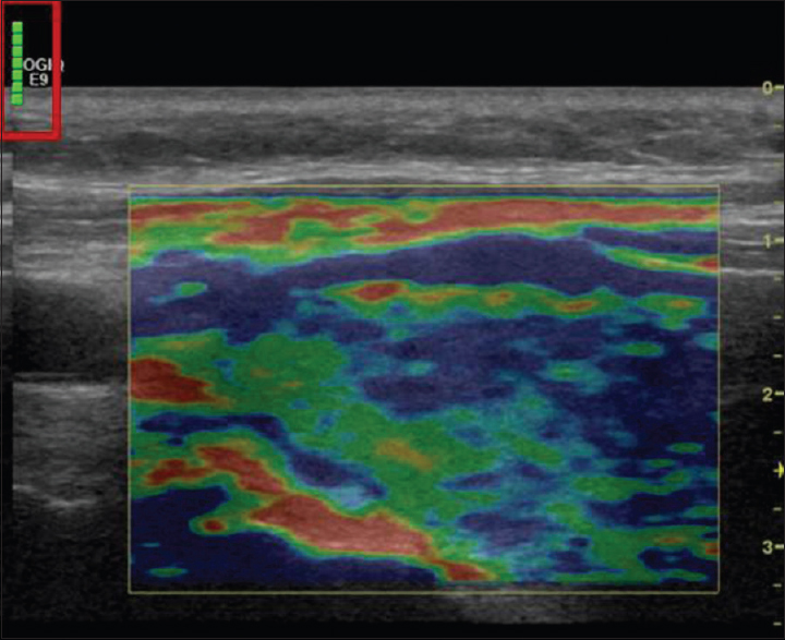
- Elastogram of right thyroid lobe 3.2 cm solid nodule. Real-time applied transducer pressure analyzed by numeric scale displayed at the right upper corner of the elastogram image (red box).
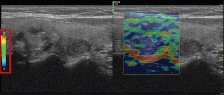
- Elastogram of right thyroid lobe upper pole 2.1 cm nodule with microcalcifications. Nodule strain elastogram is displayed by a 256-color mapping scale with red representing the softest components and blue representing the hardest components.
We separated patients into two groups, based if an elastogram was available for the radiologist to review before FNA or not. All FNA were completed by one of two fellowship-trained radiologists with specialty in ultrasound-guided procedures. A 21-gauge needle was used in every case, and aspiration was aided by a 5-cc syringe. All pathological analyses were performed by one of three board-certified cytopathologists. In all cases, the cytopathologist was present during the procedure for rapid onsite evaluation of specimen adequacy.
The final pathological diagnosis was reviewed through the institution's electronic medical record for every case. Pathology reports were available for 221 out of the 225 specimens reviewed. Adequacy was determined using the Bethesda criteria as above.
The Chi-square test was used to determine statistically significant variances in the demographic parameters. SAS 9.3 software (SAS Institute, Inc. Cary, NC) was used for calculations. P < 0.05 value was considered statistically significant. The overall ND rates were also measured.
Results
A total of 221 patients were included in the final analysis. Elastography was performed on 140 cases (63.4%). There was no significant difference between the two groups in demographics (including gender, age, and which thyroid gland contained the nodule) as well as nodule characteristics.
Regarding the specimen adequacy, the total percentage of ND aspirates was 12.2%, slightly higher than the 10% supported by the ASCP. In analyzing the two groups, when an elastogram was available before FNA, the ND rate was 10% (14/140) which was in concordance with the ASCP recommendations. However, the group without an elastogram available had an ND rate of 16% (13/81). However, this difference was not found to be statistically significant, P = 0.205.
Discussion
Thyroid nodules continue to pose a diagnostic dilemma secondary to poor sensitivity and specificity using gray scale features alone. Although considered the gold standard for nodule evaluation, FNA carries its own risks including patient pain, cost, and possible complications (although rare). The need for repeat FNA secondary to ND results can cause further patient harm as well as delay time to treatment and increase patient anxiety.
To help increase the rate of adequate specimen recovery, the use of ultrasound to guide FNA has become the standard of care. With the assistance of ultrasound-guided FNA, the rates of ND specimen aspirates still range up to 20%.[2] In a recent meta-analysis, even the addition of rapid onsite evaluation by a cytopathologist has shown considerable variability in helping to reduce ND rates.[6]
Ex vivo studies have found that malignant thyroid lesions have a stiffer architecture compared to benign thyroid stroma.[7] First described in 1991 by Ophir, tissue stiffness could be measured by applying an external axial stimulus and viewing the associated compressibility.[8] They ultimately coined the resultant map of strain profiles, an elastogram. The continued research in elastography has focused on its ability to distinguish benign and malignant lesions by “virtual palpation.”
Several studies have been performed to evaluate the role of elastography in targeting interventions. Most papers have focused on ultrasound-guided transrectal biopsies of the prostate gland. Using elastograms, small studies have been able to show enhanced detection rates as well as decreasing the number of core specimens required for diagnosis.[34] To date, there has been only one study performed evaluating strain elastography-guided FNA of thyroid nodules. In 2013, Yildrim et al., prospectively analyzed the rates of ND aspirates between specimens obtained from regions of higher strain and lower strain from 96 patients.[9] The authors found a significant improvement in specimen adequacy when aspirates were obtained from regions that demonstrate a stiff elastogram as described previously.
Our study was aimed at replicating Yildrim's results in a larger patient sample and hoping to show an improved rate of ND aspirates. In our retrospective review, although there was a decreased rate of ND specimens in the group that USE was performed (16% vs. 10%), this was not found to be statistically significant. This may be partially due to the study being underpowered to detect true significance.
Our study did have several limitations that may have also contributed to our results. First of all, we performed a retrospective review, which by its very nature allows for confounding bias. Furthermore, although an elastogram was available for the radiologist before FNA in the elastography group, we were unable to prove that stiffer regions were actually targeted and aspirated during the procedure. Furthermore, it is possible that stiffer regions of nodules were sampled in the group without elastograms even though elastography was not performed. Finally, as our study was a retrospective review, the pathologist performing the specimen analysis was not controlled for, leading to possible variability.
Another possibility exists to explain why the presence of an elastogram prior the FNA did not significantly reduce the rate of ND results. Our study was predicated on the idea that stiffer regions within nodules would likely decrease ND aspirates as these regions may represent more cellular areas. However, the development of a stiff consistency in many cancers is secondary to a desmoplastic transformation that takes place. This process is composed of activated fibroblasts (also known as myofibroblasts), tumor vessels, inflammatory cells, and extracellular matrix components. Activated carcinoma induced fibroblasts, along with other extracellular matrix proteins, comprise a dominant proportion of several carcinomas, including more than 90% of overall tumor mass in pancreatic cancers.[10] It has even been shown that the complex relationship between these components may promote tumor cell growth.[11] It is possible that the stiffer regions within thyroid nodules are more a product of fibroblastic growth than increased cellular density. This theory would not be able to explain Yildrim's results though.
Conclusions
We were unable to demonstrate a significant difference in thyroid nodule aspirate adequacy between patients who had an elastogram present before FNA compared with those without one. These results may be secondary to lack of power, poor study design versus fibroblastic growth as described above. We hope our study raises awareness of the exciting prospect of USE-targeted biopsy and springs future studies confirming its potential.
Financial support and sponsorship
Nil.
Conflicts of interest
There are no conflicts of interest.
Available FREE in open access from: http://www.clinicalimagingscience.org/text.asp?2017/7/1/4/199054.
References
- Systematic review of cases reporting blood extravasation-related complications after thyroid fine needle biopsy. J Otolaryngol Head Neck Surg. 2010;39:532-41.
- [Google Scholar]
- The Bethesda system for reporting thyroid cytopathology. Am J Clin Pathol. 2009;132:658-65.
- [Google Scholar]
- Initial experiences with real-time elastography guided biopsies of the prostate. J Urol. 2005;174:115-7.
- [Google Scholar]
- Real-time elastography for detecting prostate cancer: Preliminary experience. BJU Int. 2007;100:42-6.
- [Google Scholar]
- Management of thyroid nodules detected at US: Society of radiologists in ultrasound consensus conference statement. Radiology. 2005;237:794-800.
- [Google Scholar]
- The influence of rapid onsite evaluation on the adequacy rate of fine-needle aspiration cytology: A systematic review and meta-analysis. Am J Clin Pathol. 2013;139:300-8.
- [Google Scholar]
- Elastic moduli of thyroid tissues under compression. Ultrason Imaging. 2005;27:101-10.
- [Google Scholar]
- Elastography: A quantitative method for imaging the elasticity of biological tissues. Ultrason Imaging. 1991;13:111-34.
- [Google Scholar]
- The value of ultrasound elastography-guided fine-needle aspiration biopsy of thyroid nodules in reducing nondiagnostic results. J Med Ultrason. 2013;40:169-74.
- [Google Scholar]
- Desmoplasia of pancreatic ductal adenocarcinoma. Clin Gastroenterol Hepatol. 2009;7(11 Suppl):S44-7.
- [Google Scholar]
- Physiological mechanisms of tumor-cell invasion and migration. Physiology (Bethesda). 2005;20:194-200.
- [Google Scholar]


