Translate this page into:
Computed Tomography Angiography with a 192-slice Dual-source Computed Tomography System: Improvements in Image Quality and Radiation Dose
Address for correspondence: Philip V M Linsen, Department of Radiology, Erasmus Medical Center, CA-207, s-Gravendijkwal 230, 3015 CE, Rotterdam, The Netherlands. E-mail: philiplinsen@hotmail.com
-
Received: ,
Accepted: ,
This is an open access article distributed under the terms of the Creative Commons Attribution-NonCommercial-ShareAlike 3.0 License, which allows others to remix, tweak, and build upon the work non-commercially, as long as the author is credited and the new creations are licensed under the identical terms.
This article was originally published by Medknow Publications & Media Pvt Ltd and was migrated to Scientific Scholar after the change of Publisher.
Abstract
Purpose:
This study aims to compare image quality, radiation dose, and the influence of the heart rate on image quality of high-pitch spiral coronary computed tomography angiography (CCTA) using 128-slice (second generation) dual-source CT (DSCT) and a 192-slice DSCT (third generation) scanner.
Materials and Methods:
Two consecutive cohorts of fifty patients underwent CCTA by high-pitch spiral scan mode using 128 or 192-slice DSCT. The 192-slice DSCT system has a more powerful roentgen tube (2 × 120 kW) that allows CCTA acquisition at lower tube voltages, wider longitudinal coverage for faster table speed (732 m/s), and the use of iterative reconstruction. Objective image quality was measured as the signal-to-noise ratio (SNR) and contrast-to-noise ratio (CNR). Subjective image quality was evaluated using a Likert scale.
Results:
While the effective dose was lower with 192-slice DSCT (1.2 ± 0.5 vs. 0.6 ± 0.3 mSv; P < 0.001), the SNR (18.9 ± 4.3 vs. 11.0 ± 2.9; P < 0.001) and CNR (23.5 ± 4.8 vs. 14.3 ± 4.1; P < 0.001) were superior to 128-slice DSCT. Although patients scanned with 192-slice DSCT had a faster heart rate (59 ± 7 vs. 56 ± 6; P = 0.045), subjective image quality was scored higher (4.2 ± 0.8 vs. 3.0 ± 0.7; P < 0.001) compared to 128-slice DSCT.
Conclusions:
High-pitch spiral CCTA by 192-slice DSCT provides better image quality, despite a higher average heart rate, at lower radiation doses compared to 128-slice DSCT.
Keywords
Computed tomography
coronary angiography
heart rate
image quality
radiation dose

INTRODUCTION
Due to its high sensitivity and negative predictive value, coronary computed tomography angiography (CCTA) has emerged as a reliable noninvasive examination to rule out coronary artery disease (CAD).[12] However, exposure to radiation has remained an issue of concern.[34] Over the past decade, technical innovations have decreased the radiation dose associated with CCTA, while maintaining good diagnostic performance.[56]
For the second-generation 128-slice dual-source CT (DSCT) systems, a prospectively electrocardiogram (ECG)-triggered high-pitch spiral scan mode was introduced by which the entire heart could be scanned within the time of a single heart cycle. While the detector collimation does not completely cover the heart for a stationary table position, the entire heart can still be scanned by accelerating the spiral pitch (table advancement) and extending the exposure window during the single-beat acquisition. Previous studies have demonstrated that diagnostic image quality can be achieved at much lower radiation doses, particularly in patients with low heart rate.[789]
The 192-slice (third generation) DSCT system offers several technical improvements such as faster gantry rotation speed (from 280 to 250 ms), increased longitudinal detector coverage from collimation coverage (from 38 mm for 128-slice DSCT to 58 mm for 192-slice DSCT), and a more powerful roentgen tube (2 × 100 kW for 128-slice DSCT vs. 2 × 120 kW for 192-slice DSCT) that allows CCTA acquisition at tube voltages down to 70 kV. These improvements may allow for improved image quality, even at higher heart rates, while reducing radiation dose, when performing high-pitch spiral CCTA by 192-slice DSCT. This has been shown in ex vivo studies by Morsbach et al.[10]
In this study, we investigated the differences in image quality, radiation dose, and the influence of the heart rate on image quality in high-pitch spiral CCTA performed by 128 and 192-slice DSCT.
MATERIALS AND METHODS
Study design
For both 128 and 192-slice DSCT, the first, consecutive fifty patients scanned using a high-pitch spiral mode with known or suspect CAD were identified from medical records. Exclusion criteria were age younger than 18 years old, previous coronary artery bypass graft, and the inability to follow instructions needed for the CCTA. In case of second-attempt scans because of nondiagnostic image quality, only the first scan was included.
The 128-slice DSCT scans were performed between 28 April 2009 and 2 September 2009. The 192-slice DSCT scans were acquired between 3 March 2014 and 17 December 2014. According to institutional standard protocols, a noncontrast calcium scan was performed before CCTA. The calcium score was evaluated using dedicated software (CA Scoring; Siemens Medical Solutions, Forchheim, Germany) and expressed as the Agatston score.[11] The Institutional Review Board approved the study. Given the retrospective nature of the study, no informed consent was required.
Computed tomography acquisition
All patients received sublingual nitroglycerin before the CCTA examination. Intravenous beta-blockers were administered in patients with higher heart rates. Data acquisition was prospectively ECG-triggered to start at 65% of the R-R interval and completed within one cardiac cycle. All contrast-enhanced CCTA data were reconstructed with a slice thickness of 0.75 mm and slice increment of 0.3 mm.
The 128-slice DSCT system (Somatom Definition Flash, Siemens Medical Solutions, Forchheim, Germany) had a collimation of 2 × 128 × 0.6 mm, using a flying focal spot technique and a gantry rotation time of 280 ms.[12] The high-pitch spiral acquisition was made with a fixed pitch of 3.4 corresponding to a table movement of 4.58 m/s. Tube voltage (100 or 120 kV) was selected manually, and automatic exposure control (AEC) was used for the tube current. Images were reconstructed using filtered back projection and a medium-smooth kernel (B26f).
The 192-slice DSCT system (Somatom Force, Siemens Medical Solutions, Forchheim, Germany) had a collimation of 2 × 192 × 0.6 mm, using a flying focal spot technique and a gantry rotation time of 250 ms. The high-pitch spiral acquisition was made with a fixed pitch of 3.2 corresponding to a table movement of 7.37 m/s. Tube voltage was selected semi-automatically by the automatic selection algorithm, and AEC was used for the tube current. With 192-slice DSCT, the CCTA acquisition was possible at tube voltage levels between 70 and 120 kV in steps of 10 kV. Slices were reconstructed using a medium sharp kernel (Bv40), using model-based iterative reconstruction strength level 3 (ADMIRE; Siemens Medical Solutions, Forchheim, Germany).
Contrast protocols
The 128-slice DSCT protocol applied a bolus-tracking protocol. CCTA acquisition began once a threshold of 74 HU was exceeded within the ascending aorta. Timing of the scan using the 192-slice DSCT system was determined using a test bolus, planning the acquisition 8 s after peak enhancement of the test bolus. Most patients (94) were examined using iopromide 370 mg/l (Ultravist 370; Bayer Healthcare, Berlin, Germany), whereas six patients were examined using iodixanol 320 mg/l (Visipaque 320; GE Healthcare, Milwaukee, USA). For simplicity, the latter six patients (all in the 192-slice DSCT group) were not included in the contrast delivery comparison. The contrast injection was followed by a 45 ml saline chaser.
Objective image quality measurements
Both objective and subjective image quality was assessed using a dedicated workstation (Syngo.via, Siemens Medical Solutions, Forchheim, Germany). The objective image quality was assessed using the signal-to-noise ratio (SNR) and contrast-to-noise ratio (CNR). Segments 1, 2, 3, 5, 6, 7, 8, 11, 13, and 15 were analyzed, in accordance with the coronary artery model of the Society of Cardiovascular Computed Tomography.[13]
Circular regions of interest (ROI), as large as possible, were placed in each coronary segment by one observer. The vessel wall, calcifications, plaques, or stents were carefully avoided. Coronary segments without interpretable lumen due to excessive plaque were excluded from the analysis. For the mean pericardial fat values, two samples near the right and left coronary artery were averaged. To calculate the SNR and CNR, the following formulas were used:[14]
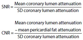
The standard deviation (SD) of each ROI represented the image noise.[15] The CNR was calculated for the proximal segments 1, 5, 6, and 11, following the methodology by Achenbach et al.[16]
Subjective image quality
The subjective image quality was assessed independently by two observers blinded to the type of scanner and any other technical or medical information. A five-point Likert scale was used to score the image quality. The following scores were possible: 1 - poor, impaired image quality limited by excessive noise or poor vessel wall definition; 2 - adequate, reduced image quality either poor vessel wall definition or excessive image noise, limitation in low contrast resolution remain evident; 3 - good, effect of image noise, limitations of low contrast resolution, and vessel margin definition are minimal; 4 - very good, good attenuation of vessel lumen and delineation of vessel walls, relative image noise is minimal, coronary wall definition and low contrast resolution well maintained; 5 - excellent, excellent attenuation of the vessel lumen and clear delineation of vessel walls, limited perceived image noise.[17] For the final analysis of image quality in relation to acquisition properties, a consensus reading was performed for all segments with discordant scores beyond one point.
Radiation dose
The radiation dose was reported as volume CT dose index (CTDIvol), dose-length product (DLP), and effective dose (ED). For each patient, the CTDIvol was recorded from the automatically generated patient protocol. The estimated ED was calculated with the formula DLP × 0.014, using the 0.014 conversion factor for chest radiation (in mSv/Gy/cm) according to the European Guidelines for Multislice Computed Tomography and as adopted in large trials.[1819]
Statistical analyses
Continuous variables were expressed as mean ± SD or median (range) as appropriate, and categorical variables as frequencies or percentages. Student's t-test or Mann–Whitney U-test was used to compare the patient's characteristics, the amount and infusion rate of contrast, SNR, CNR, and radiation dose. Risk factors were analyzed using Chi-square test. To compare the subjective image quality, Mann–Whitney U-test was used. The impact of mean heart rate on image quality was assessed by Spearman's rank correlation coefficient. A two-tailed P < 0.05 was considered significant. The statistical analyses were performed with SPSS (version 21.0, SPSS Inc., Chicago, IL, USA).
RESULTS
Study population
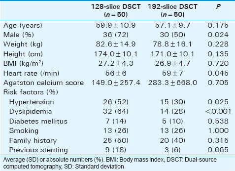
The mean age in the 192-slice DSCT group was 57.1 ± 9.7 compared to 59.9 ± 10.9 in 128-slice DSCT group (P = 0.175). The mean heart rate during acquisition was higher for 192-slice DSCT (59 ± 7) compared with 128-slice DSCT (56 ± 6; P = 0.045) [Table 1]. In the 192-slice DSCT group, eight patients were rescanned due to movement/breathing artifacts (7) or an improperly set scanning coverage (1). Eleven patients were rescanned in the 128-slice DSCT group due to movement/breathing artifacts (9) or inadequate contrast timing (2) (P = 0.447). The second attempt scans were excluded from analysis.
Objective image quality
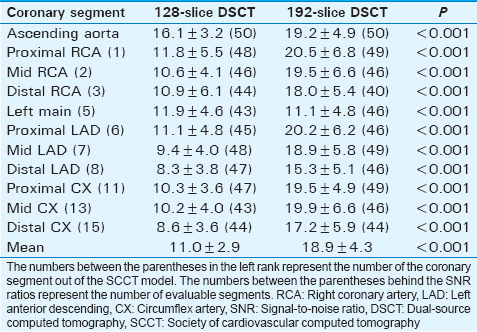
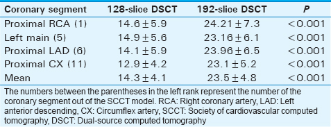
Mean coronary lumen attenuation was 572 HU for 192-slice DSCT and 401 HU for 128-slice DSCT. Mean subcutaneous fat attenuation was 123 HU for 192-slice DSCT and 107 HU for 128-slice DSCT. As seen in Table 2, the mean SNR for scans made by 192-slice was higher (18.9 ± 4.3) than that for scans made by 128-slice DSCT (11.0 ± 2.9; P < 0.001). Table 3 shows that the mean CNR was higher for 192-slice DSCT (23.5 ± 4.8) compared to 128-slice DSCT (14.3 ± 4.1; P < 0.001).
Subjective image quality
Out of 500 potentially available coronary segments, 463 were evaluable for the 192-slice DSCT group and 454 in the 128-slice DSCT group (P = 0.302). The mean subjective image quality score for the 192-slice DSCT group was 4.2 ± 0.8 compared to 3.0 ± 0.7 in the 128-slice DSCT group (P < 0.001). Individual coronary segment scores were better on scans performed by 192-slice DSCT compared to 128-slice DSCT [Table 4].
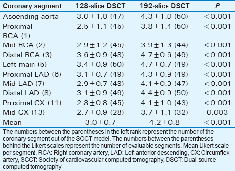
Impact of heart rate on image quality
There was a borderline significant correlation between low heart rate and mean image quality score on a per-patient analysis 128-slice DSCT (r = −0.278; P = 0.051) and less evident for 192-slice DSCT (r = −0.164; P = 0.265) [Figure 1].
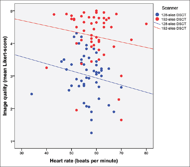
- The Influence of heart rate on image quality. It shows the linear regression plot of mean image quality scores overall coronary segments per patient (y-axis) against heart rate during computed tomography scanning (x-axis) in 128-slice dual-source computed tomography and 192-slice dual-source computed tomography. The dots represent the individual patients. The lines represent the Spearman's rank correlation coefficient. The lines show that image quality will decrease with higher heart rate. It also shows that 192-slice dual-source computed tomography is superior to 128-slice dual-source computed tomography in coronary computed tomography angiography at all heart rates and maintains good diagnostic image quality at higher heart rates.
Contrast
By including only patients scanned using iopromide 370 mg/l, the mean amount of contrast was lower for 192-slice DSCT (64.3 ± 4.1 ml compared to 75.6 ± 6.1; P < 0.001). The average infusion speed was 5.4 ± 0.2 ml/s for 192-slice DSCT and 5.9 ± 0.3 for 128-slice DSCT (P < 0.001).
Radiation dose
The radiation dose using 192-slice DSCT was lower than that at 128-slice DSCT (0.6 ± 0.3 mSv vs. 1.2 ± 0.5 mSv; P < 0.001). The tube voltage used was lower for the 192-slice DSCT, as 17 scans were made at 70 kV. The scan length was not significantly different [Table 5].

DISCUSSION
The main findings of this paper are that high-pitch spiral CCTA by 192-slice DSCT combined with iterative reconstruction is associated with better image quality and lower exposure to radiation and contrast medium, compared with 128-slice DSCT.
CCTA has developed as a reliable noninvasive diagnostic tool to assess CAD. In the recent years, there have been numerous technological improvements in CT technology to reduce radiation exposure while maintaining high image quality. As confirmed in our study, the 192-slice DSCT system allows for a further reduction in radiation dose.[81020] With the latest 192-slice DSCT system, it is possible to perform CCTA with a radiation exposure far below 1 mSv in most of the patients. An example of good diagnostic quality imaging at 192-slice DSCT can be found in Figure 2.
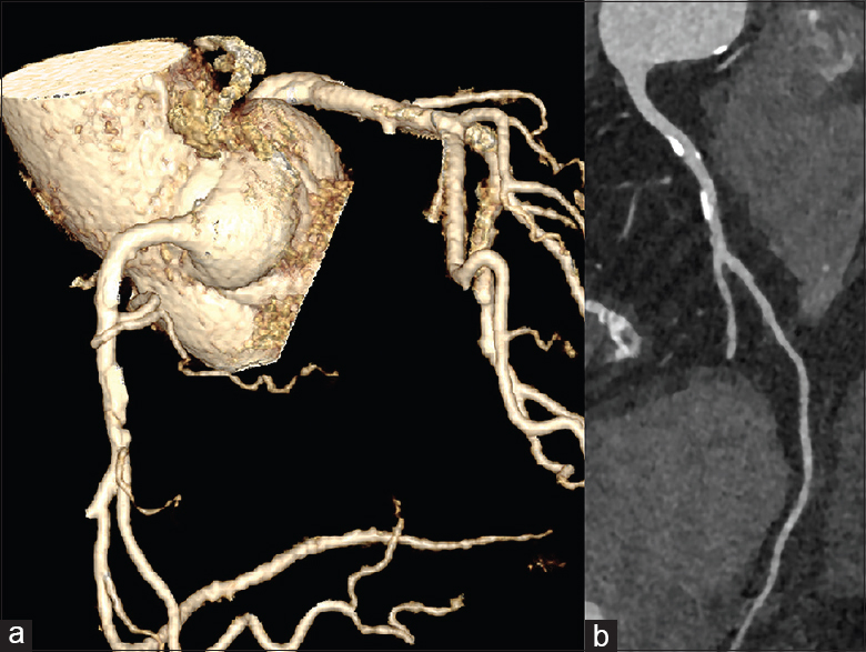
- A 67-year old female with symptoms related to angina and an Agatston score of 632, a BMI of 24.3 kg/m2, and a heart rate during scanning of 67 beats per minute. (a) An example of a volume rendered multiplanar reconstruction of the right coronary artery (RCA) from a coronary computed tomography angiography performed at 192-slice dual-source computed tomography using a tube voltage of only 70 kV. (b) The same RCA in curved multiplanar reconstruction. Both demonstrate superior diagnostic image quality despite the low kV settings.
As seen in our study, radiation exposure was lowered by approximately 50% comparing 192- and 128-slice DSCT. This was partly because with 192-slice DSCT, a more powerful roentgen tube became available. This allowed CCTA at 70 kV rather than 100 kV at 128-slice DSCT.
The improvement in image quality with the 192-slice DSCT system is multifactorial. Faster rotation speed and wider coverage lower the sensitivity for motion artifacts. At lower tube voltage, the photoelectronic effect of iodine results in a higher signal and improved contrast with surrounding tissues. While on the other hand, the higher tube current avoids image noise, also iterative reconstruction algorithms contributed to improved SNR and CNR with 192-slice DSCT. In our study, we used a mean of 64 ml iodine contrast while contrast medium volumes in similar studies ranged from 10 to around 60 ml using 80 kV as the lowest tube voltage.[2122]
Similar to the findings by Ghadri et al., we did not observe a significant correlation between heart rate and image quality while using the high-pitch spiral scan mode, particularly for the 192-slice DSCT system.[23] There seems to be a negative tendency but, like the study by Ghadri et al., most likely this study is insufficiently powered to show a significant correlation.
Our results have to be evaluated in light of some limitations. The study is based on a historical comparison, and there is no guarantee that the populations are entirely comparable. In addition, experience with the performance of high-pitch spiral CCTA has increased over time. Improvement of image quality may be multifactorial, and not just the result of improved scanner hardware. Because the difference in attenuation between iodine and other tissues increases at lower kV tube setting, the attenuation values in the coronary lumen were higher for 192-slice DSCT despite the lower amounts of administrated contrast medium. The more robust capabilities for high-quality low tube voltage scanning in a less strictly selected population may bode well for the routine implementation of high-pitch spiral CCTA.
Furthermore, iterative reconstruction techniques were not yet available when 128-slice DSCT was introduced at our center. The use of iterative reconstruction for the 192-slice DSCT system further improved the SNR and CNR using algorithms which permit a decoupling of spatial resolution and noise.[242526]
CONCLUSIONS
High-pitch spiral CCTA on the 192-slice DSCT system results in higher image quality while the radiation dose is further reduced compared to 128-slice DSCT. The 192-slice DSCT system also allows for the use of the high-pitch spiral scan mode in patients with higher heart rates while maintaining good diagnostic image quality.
Financial support and sponsorship
Nil.
Conflicts of interest
Marcel L. Dijkshoorn: Consultant Siemens Medical Solutions. Koen Nieman: Institutional Research support from Bayer Healthcare, GE Healthcare, Siemens Medical Solutions.
Available FREE in open access from: http://www.clinicalimagingscience.org/text.asp?2016/6/1/44/192840
REFERENCES
- Diagnostic accuracy of 64-slice computed tomography coronary angiography: A prospective, multicenter, multivendor study. J Am Coll Cardiol. 2008;52:2135-44.
- [Google Scholar]
- Low-dose coronary computed tomography angiography using prospective ECG-triggering compared to invasive coronary angiography. Int J Cardiovasc Imaging. 2011;27:425-31.
- [Google Scholar]
- Estimating risk of cancer associated with radiation exposure from 64-slice computed tomography coronary angiography. JAMA. 2007;298:317-23.
- [Google Scholar]
- Radiation dose of cardiac computed tomography – What has been achieved and what needs to be done. Eur Radiol. 2011;21:505-9.
- [Google Scholar]
- Effect of Tube voltage (100 vs 120 kVp) on radiation dose and image quality using prospective gating 320 row multi-detector computed tomography angiography. J Clin Imaging Sci. 2013;3:62.
- [Google Scholar]
- Coronary CT angiography of patients with a normal body mass index using 80 kVp versus 100 kVp: A prospective, multicenter, multivendor randomized trial. AJR Am J Roentgenol. 2011;197:W860-7.
- [Google Scholar]
- High-pitch coronary CT angiography in dual-source CT during free breathing vs. breath holding in patients with low heart rates. Eur J Radiol. 2013;82:2217-21.
- [Google Scholar]
- Closing in on the K edge: Coronary CT angiography at 100, 80, and 70 kV-initial comparison of a second- versus a third-generation dual-source CT system. Radiology. 2014;273:373-82.
- [Google Scholar]
- Image quality and radiation dose of a prospectively electrocardiography-triggered high-pitch data acquisition strategy for coronary CT angiography: The multicenter, randomized PROTECTION IV study. J Cardiovasc Comput Tomogr. 2015;9:278-85.
- [Google Scholar]
- Performance of turbo high-pitch dual-source CT for coronary CT angiography: First ex vivo and patient experience. Eur Radiol. 2014;24:1889-95.
- [Google Scholar]
- Quantification of coronary artery calcium using ultrafast computed tomography. J Am Coll Cardiol. 1990;15:827-32.
- [Google Scholar]
- Image reconstruction and image quality evaluation for a 64-slice CT scanner with z-flying focal spot. Med Phys. 2005;32:2536-47.
- [Google Scholar]
- SCCT guidelines for the interpretation and reporting of coronary CT angiography: A report of the Society of Cardiovascular Computed Tomography Guidelines Committee. J Cardiovasc Comput Tomogr. 2014;8:342-58.
- [Google Scholar]
- Influence of convolution filtering on coronary plaque attenuation values: Observations in an ex vivo model of multislice computed tomography coronary angiography. Eur Radiol. 2007;17:1842-9.
- [Google Scholar]
- Impact on image quality and radiation exposure in coronary CT angiography: 100 kVp versus 120 kVp. Acta Radiol. 2010;51:903-9.
- [Google Scholar]
- Comparison of image quality in contrast-enhanced coronary-artery visualization by electron beam tomography and retrospectively electrocardiogram-gated multislice spiral computed tomography. Invest Radiol. 2003;38:119-28.
- [Google Scholar]
- Adaptive statistical iterative reconstruction: Assessment of image noise and image quality in coronary CT angiography. AJR Am J Roentgenol. 2010;195:649-54.
- [Google Scholar]
- The 2007 recommendations of the international commission on radiological protection. ICRP publication 103. Ann ICRP. 2007;37:1-332.
- [Google Scholar]
- Estimated radiation dose associated with cardiac CT angiography. JAMA. 2009;301:500-7.
- [Google Scholar]
- Prospectively ECG-triggered high-pitch coronary angiography with third-generation dual-source CT at 70 kVp tube voltage: Feasibility, image quality, radiation dose, and effect of iterative reconstruction. J Cardiovasc Comput Tomogr. 2014;8:418-25.
- [Google Scholar]
- Image quality of ultra-low radiation exposure coronary CT angiography with an effective dose <0.1 mSv using high-pitch spiral acquisition and raw data-based iterative reconstruction. Eur Radiol. 2013;23:597-606.
- [Google Scholar]
- Coronary computed tomography angiography using ultra-low-dose contrast media: Radiation dose and image quality. Int J Cardiovasc Imaging. 2013;29:1335-40.
- [Google Scholar]
- Image quality and radiation dose comparison of prospectively triggered low-dose CCTA: 128-slice dual-source high-pitch spiral versus 64-slice single-source sequential acquisition. Int J Cardiovasc Imaging. 2012;28:1217-25.
- [Google Scholar]
- Optimizing radiation dose by using advanced modelled iterative reconstruction in high-pitch coronary CT angiography. Eur Radiol. 2016;26:459-68.
- [Google Scholar]
- Detection of coronary artery stenosis with sub-milliSievert radiation dose by prospectively ECG-triggered high-pitch spiral CT angiography and iterative reconstruction. Eur Radiol. 2013;23:2927-33.
- [Google Scholar]
- Chest computed tomography using iterative reconstruction vs filtered back projection (Part 1): Evaluation of image noise reduction in 32 patients. Eur Radiol. 2011;21:627-35.
- [Google Scholar]


