Translate this page into:
Basal Cell Adenocarcinoma of the Minor Salivary Glands Involving Palate and Maxillary Sinus
Address for correspondence: Dr. Prathi Venkata Sarath, Department of Oral Medicine and Radiology, Narayana Dental College and Hospital, Chinthareddypalem, Nellore, Andhra Pradesh, India. E-mail: sarathprathi@gmail.com
-
Received: ,
Accepted: ,
This is an open-access article distributed under the terms of the Creative Commons Attribution License, which permits unrestricted use, distribution, and reproduction in any medium, provided the original author and source are credited.
This article was originally published by Medknow Publications & Media Pvt Ltd and was migrated to Scientific Scholar after the change of Publisher.
Abstract
Basal cell adenocarcinoma (BCAC) is a rare neoplasm accounting for only 2.9% of all salivary gland neoplasms. BCAC involving palatal minor salivary glands are exceedingly rare, and only 10 cases have been reported in the literature. The treatment of choice is surgical excision. Here, we report a case of a 55-year-old male patient with massive BCAC of palatal minor salivary gland extending into the maxillary sinus. This is the first case of BCAC treated by radiotherapy followed by chemotherapy. A follow-up check conducted after 14-months showed good prognosis.
Keywords
Basal cell adenocarcinoma
minor salivary glands
salivary gland tumor
INTRODUCTION

In 2005, World Health Organization defined Basal cell adenocarcinoma (BCAC) as “an epithelial neoplasm that has cytological characteristics of Basal cell adenoma (BCA), but a morphologic growth pattern indicative of malignancy.”[1] BCAC is classified as a low-grade malignancy predominantly affecting parotid and submandibular salivary glands. It is considered as the malignant counterpart of the BCA. Histological differentiation between the two is difficult, and they are often discriminated only by the invasion of local structures or by Perineural or vascular invasion. It is necessary to differentiate BCAC from other basaloid cell tumors of the minor salivary glands such as canalicular adenoma, BCA, adenoid cystic carcinoma, polymorphous low-grade adenocarcinoma, myoepthelial tumors, epithelial-myoepthelial carcinoma, and basaloid squamous cell carcinoma because of the prognosis and potential differences in the treatment.[2] Surgical excision is the primary treatment of choice.
CASE REPORT
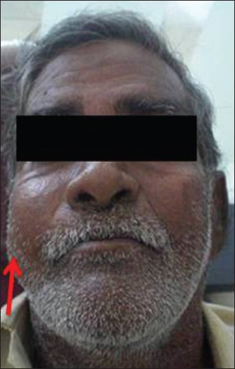
A 55-year-old male patient presented with a complaint of pain and swelling in the right side of the palate that had persisted for a month. The patient revealed that pain was mild, throbbing, and got aggravated while eating. The swelling was gradually increasing in size. Extra-oral examination revealed soft, tender, and diffuse swelling in the right middle third of the face measuring approximately about about 4cm × 4cm, superioinferiorly extending from the infraorbital region to 1 cm above the right commissure of the lip and anteroposteriorly from the right nasolabial fold to 2 cm anterior to the tragus [Figure 1]. Right submandibular lymph node was enlarged and tender on palpation. Intra-oral examination revealed the presence of right palatal swelling measuring approximately 4 cm × 2.5 cm, which was soft to firm in consistency and tender on palpation [Figure 2]. On correlating the history and clinical examination, this case was provisionally diagnosed as pleomorphic adenoma of the palatal minor salivary glands.

- 55-year-old male patient with pain and swelling of the palate which was subsequently diagnosed as adenoma of the palatal minor salivary glands. Extra-oral clinical photograph shows swelling on the right side of the face.

- 55-year-old male patient with pain and swelling of the palate which was subsequently diagnosed as adenoma of the palatal minor salivary glands. Intra-oral clinical photograph reveals swelling on the right side of the palate.
Radiographic examination of the para nasal sinus revealed the presence of a diffuse radio-opaque mass occupying entire right maxillary sinus without involving the orbit [Figure 3]. Enhanced computed tomography with slice thickness of 4 mm showed a homogenous mass infiltrating the palate, nasal cavity, and right maxillary sinus measuring approximately about 4 cm × 4 cm superioinferiorly and 3 cm × 2 cm anteroposteriorly [Figure 4]. Nasopharayngoscopy revealed lateral wall of the right nasal cavity was pushed medially by the mass. Routine hematological investigations such as hemoglobin (12.5 g/dl), red blood cell count (4.0 million/cu.mm), total white blood cell count (12,200), differential leucocyte count (Neutrophils-77%, lymphocytes-15%, monocytes-3%, eosinophils-5%), and platelet count (1.9 lakhs/cu.mm) Were within the normal limits. Under local anesthesia, a biopsy from the palatal region was taken. Multiple grayish white bits of soft-tissue measuring approximately about 1.0 cm × 0.5 cm × 0.4 cm were obtained. Histopathological section showed nests, cords, and trabecular of small dark staining cells with variably sized vesicular or hyper chromatic nuclei. Occasional ductal structures and mitotic figures were noticed. Frank infiltration into the deeper connective tissue, focal areas of necrosis, and hemorrhage were also seen [Figures 5 and 6]. Histopathological features were consistent with poorly differentiated malignancy. Later, immunohistochemistry showed positive reaction to pan-cytokeratin and negative to calponin and p63 suggestive of BCAC. Age of the patient and size of the tumor precluded surgical management of the lesion; hence, patient was subjected to radiotherapy followed by chemotherapy. Post-treatment patient's course was uneventful and there have been no signs of local recurrence or distant metastasis during the 14-month follow-up [Figures 7 and 8].

- 55-year-old male patient with pain and swelling of the palate which was subsequently diagnosed as adenoma of the palatal minor salivary glands. X-ray of the paranasal sinus shows diffuse radiopaque mass (arrow) involving right maxillary sinus.

- 55-year-old male patient with pain and swelling of the palate which was subsequently diagnosed as adenoma of the palatal minor salivary glands. Enhanced computed tomography demonstrates homogenous mass (arrow) in the right middle third of the face involving the nasal cavity and maxillary sinus.

- 55-year-old male patient with pain and swelling of the palate which was subsequently diagnosed as adenoma of the palatal minor salivary glands. Hematoxylin and eosin stained palatal tissue (×100) shows cords (black arrow), islands of basaloid cells (yellow arrow) in close proximity to overlying hyperplastic palatal epithelium and also infi ltrating into deeper connective tissue.

- 55-year-old male patient with pain and swelling of the palate which was subsequently diagnosed as adenoma of the palatal minor salivary glands. Hematoxylin and eosin stained palatal tissue (×40) reveals aggregates of basaloid cells, with pleomorphic vesicular or hyper chromatic nuclei (yellow arrow) with occasional ductal structures (black arrow).

- 55-year-old male patient with pain and swelling of the palate which was subsequently diagnosed as adenoma of the palatal minor salivary glands. Post-treatment extra-oral clinical photograph shows complete resolution of the swelling on the right side of the face.

- 55-year-old male patient with pain and swelling of the palate which was subsequently diagnosed as adenoma of the palatal minor salivary glands. Post-operative intra-oral clinical photograph shows resolution of the swelling in the palate.
DISCUSSION
BCAC of palatal minor salivary glands is exceedingly rare; only 10 cases have been reported in the literature.[3] Buccal and labial minor salivary glands are also involved. BCAC typically arise in individuals older than 60 years without gender predominance and are smaller in size. The tumor grows slowly and most patients are asymptomatic.[4] In the present case, the 55-year-old male patient presented with a massive tumor (4 cm × 4 cm) extending from the infraorbital region to 1 cm above the right commissure of the lip superioinferiorly and anteroposteriorly from the right nasolabial fold to 2 cm anterior to the tragus.
BCACs consist of several types: solid, trabecular, tubular, and membranous. The solid type is more common and has high-risk of metastasis.[5] In the present case, the BCAC had a trabecular pattern, which is rare.
Cellular atypia and number of mitotic figures are greater in BCAC than in BCA. The diagnosis of BCAC may be difficult because microscopic examination of biopsy specimens sometimes is unable to distinguish it from BCA. If a tumor has a marked similarity to BCA, further examination is necessary to determine the final diagnosis. It has been reported that the examination of cell proliferation, apoptosis, and expressions of p53, bcl-2 and epidermal growth factor receptor may be useful in distinguishing malignant basal cell tumors arising in the salivary glands from their benign counterparts.[6] Here, histopathology report revealed BCAC and myoepthelial carcinoma as possible considerations. Later, positive immunoreaction to pan-cytokeratin and negative to p63 and calponin confirmed the diagnosis of BCAC.
Surgical excision with a wide margin to ensure complete removal has been recommended as the primary treatment for BCAC.[7] The local recurrence rate, metastatic rate, and mortality vary between the minor salivary glands (71%, 21% and 29% respectively) and the major salivary glands (37%, 11% and 3% respectively).[8] In the present case, the tumor infiltrated the nasal cavity and maxillary sinus. Massive size of the tumor precluded surgical management of the lesion. Hence, radiotherapy followed by adjuvant chemotherapy was preferred as a mode of treatment.[3] A 14-month follow-up of the case showed good prognosis.
CONCLUSION
BCAC of the palatal minor salivary glands is rare. Only 10 cases have been reported in the literature. In the present case, the lesion was large, infiltrating the palate, nasal cavity, and right maxillary sinus in contrast to small size lesions mentioned in the literature. Immunohistochemistry is necessary to differentiate between BCA and BCAC. Surgery is the treatment of choice, after considering the patient's age and extent of the tumor. In the present case, the patient was managed with radiotherapy followed by chemotherapy and showed good prognosis on follow-up.
ACKNOWLEDGMENT
The Author would like to thank the patient for providing consent to use his photograph in this article.
Available FREE in open access from: http://www.clinicalimagingscience.org/text.asp?2013/3/2/4/112799
Source of Support: Nil
Conflict of Interest: None declared
REFERENCES
- Pathology and genetics of tumors of the head and neck. In: World Health Organization Classification of Tumors. Vol 9. Lyon: IARC Press; 2005.
- [Google Scholar]
- Differential diagnosis of basaloid salivary gland tumors. Pathologe. 2004;25:46-55.
- [Google Scholar]
- A massive basal cell adenocarcinoma of the palatal minor salivary gland that progressed into the pterygopalatine fossa. Int J Oral Maxillofac Surg. 2012;41:444-7.
- [Google Scholar]
- Basal cell adenocarcinoma of the salivary glands.Report of seven cases and review of the literature. Cancer. 1996;78:2471-7.
- [Google Scholar]
- Pathology of salivary gland disease. In: Myers EN, Ferris RL, eds. Salivary Gland Disorder. Springer; 2007. p. :449.
- [Google Scholar]
- Basal cell adenocarcinoma of the salivary glands: Comparison with basal cell adenoma through assessment of cell proliferation, apoptosis, and expression of p53 and bcl-2. Cancer. 1998;82:439-47.
- [Google Scholar]
- Malignant epithelialtumors. In: Rosai J, Sobin LH, eds. Atlas of Tumor Pathology. 3rd Series, Fascicle 17. Tumor of Salivary Glands. Washington DC: Armed Forces Institute of Pathology; 1996. p. :257-67.
- [Google Scholar]
- Salivary gland tumors. In: Fletcher CD, ed. Diagnostic Histopathology of Tumors (2nd ed). London: Churchill Livingstone; 2000. p. :231-311.
- [Google Scholar]


