More results...

Generic selectors
Exact matches only
Search in title
Search in content
Post Type Selectors
Filter by Categories
Abdominal Radiology, Original Research
Abdominal Radiology, Pictorial Essay
Anthropology and Dental Radiology, Original Research
Breast Imaging, Case Report
Breast Imaging, Original Research
Breast Imaging, Pictorial Essay
Breast Imaging, Review Article
Cardiopulmonary Imaging, Case Report
Cardiopulmonary Imaging, Case Series
Cardiopulmonary Imaging, Education
Cardiopulmonary Imaging, Original Research
Cardiopulmonary Imaging, Pictorial Essay
Cardiopulmonary Imaging, Review Article
Case Report
Case Report, Breast Imaging
Case Report, Cardiopulmonary Imaging
Case Report, Diagnostic Radiology
Case Report, Gastrointestinal Imaging
Case Report, Genitourinary and Gynecologic Imaging
Case Report, Neuroradiology/Head and Neck Imaging
Case Report, Vascular and Interventional Radiology
Case Series
Case Series, Abdominal Radiology
Case Series, Diagnostic Radiology
Case Series, Musculoskeletal Imaging
Case Series, Vascular and Interventional Radiology
DENTAL ARTICLE
Dental Radiology, Case Report
Dental Radiology, Case Series
Dental Radiology, Original Research
Diagnostic Radiology, Case Report
Diagnostic Radiology, Case Series
Diagnostic Radiology, Original Research
Diagnostic Radiology, Pictorial Essay
Diagnostic Radiology, Review Article
Editorial
Education
Education, Diagnostic Radiology
Education, Imaging science
Education, Original Research
Emergency, Original Research
Erratum
Gastrointestinal Imaging, Case Report
Gastrointestinal Imaging, Case Series
Gastrointestinal Imaging, Original Research
Gastrointestinal Imaging, Pictorial Essay
General and Emergency Radiology, Original Research
General and Emergency Radiology, Review Article
Genitourinary and Gynecologic Imaging, Case Report
Genitourinary and Gynecologic Imaging, Original Research
Imaging Science, Original Research
Interventional Radiology, Original Research
Letter to Editor
Musculoskeletal Imaging, Case Report
Musculoskeletal Imaging, Original Article
Musculoskeletal Imaging, Original Research
Musculoskeletal Imaging, Pictorial Essay
Musculoskeletal Imaging, Review Article
Neuroradiology , Review Article
Neuroradiology Head and Neck Imaging, Pictorial Essay
Neuroradiology, Case Report
Neuroradiology, Pictorial Essay
Neuroradiology/Head and Neck Imaging, Case Report
Neuroradiology/Head and Neck Imaging, Case Series
Neuroradiology/Head and Neck Imaging, Original Research
Neuroradiology/Head and Neck Imaging, Review Article
Neuroradiology/Spine Imaging, Original Research
Notice of Retraction
Nuclear Medicine, Case Report
Nuclear Medicine, Original Research
Nuclear Medicine, Pictorial Essay
Original Article
Original Article, Neuroradiology
Original Research
Original Research Article
Original Research, Cardiopulmonary Imaging
Original Research, Dental Radiology
Original Research, Descriptive Study
Original Research, Diagnostic Radiology
Original Research, Gastrointestinal Imaging
Original Research, Genitourinary and Gynecologic Imaging
Original Research, Musculoskeletal Imaging
Original Research, Neuroradiology/Head and Neck Imaging
Original Research, Nuclear Medicine
Original Research, Vascular and Interventional Radiology
Pediatric Imaging, Case Report
Pediatric Imaging, Pictorial Essay
PICTORIAL ESSAY
Pictorial essay, Musculoskeletal Imaging
Pictorial essay, Neuroradiology/Head and Neck Imaging
PICTORIAL REVIEW
Radiologic-Pathologic Correlation
RADIOLOGICAL-PATHOLOGICAL CORRELATION
Radiology Business, Original Research
Research Article
Review Article
Review Article, Diagnostic Radiology
Review Article, Gastrointestinal imaging
Review Article, Musculoskeletal
Review Article, Musculoskeletal Imaging
Review Article, Vascular and Interventional Radiology
Technical Innovation
Technical Innovation, Cardiopulmonary Imaging
Technical Innovation, Gastrointestinal Imaging
Ultrasound, Case Report
Ultrasound, Original Research
Ultrasound, Review Article
Vascular and International Radiology, Case Report
Vascular and Interventional Radiology, Case Report
Vascular and Interventional Radiology, Case Series
Vascular and Interventional Radiology, Original Research
Vascular and Interventional Radiology, Pictorial Essay
Vascular and Interventional Radiology, Review Article
Vascular and Interventional Radiology, Short Communication
Vascular and Interventional, Original Research
View/Download PDF

Translate this page into:

CASE REPORT
2012
:2;
9
doi:
10.4103/2156-7514.94021

Asymptomatic Incidental Ductal Carcinoma in situ in a Male Breast Presenting with Contralateral Gynecomastia

Department of Radiology, Medical University of South Carolina, Charleston, SC, USA
Department of Pathology, Medical University of South Carolina, Charleston, SC, USA

Address for correspondence: Dr. Laura Isley, Department of Radiology, MSC 323, Medical University of South Carolina, 96 Jonathan Lucas Street, Rm. 210 CSB, Charleston, SC 29425-3230, USA laura.isley1@gmail.com

Licence

This is an open-access article distributed under the terms of the Creative Commons Attribution License, which permits unrestricted use, distribution, and reproduction in any medium, provided the original author and source are credited.

Disclaimer:
This article was originally published by Medknow Publications & Media Pvt Ltd and was migrated to Scientific Scholar after the change of Publisher.

Abstract

Ductal carcinoma in situ (DCIS) in males is rare and usually presents with symptoms on the affected side, such as, palpable mass or bloody nipple discharge. Even as DCIS has been reported in conjunction with gynecomastia in the same breast, we report an unusual case of a 62-year-old Caucasian male, with no family history of breast cancer, who presented with symptomatic side gynecomastia, and was incidentally found to have DCIS in a completely asymptomatic left breast. To the best of our knowledge, this case is the first report in literature of asymptomatic, incidentally discovered DCIS in a male patient.

Keywords

Ductal carcinoma in situ
male DCIS
male breast cancer

INTRODUCTION

Gynecomastia is an over-development of the glandular tissue of the breast region in males. This is different from fat deposits in the breast area seen in obese men. It occurs in young boys during puberty and in older men and results from an imbalance in the hormone levels. This is a benign condition. Breast cancer in men is uncommon. Coexistence of gynecomastia and ductal carcinoma, though rare, has been reported in literature.

CASE REPORT

A 62-year-old Caucasian male presented with a complaint of pain and swelling in the retroareolar right breast. The left breast was asymptomatic. The patient denied any family history of breast cancer and had no history of trauma to the breast. His only medication was Ritalin.

The patient underwent a bilateral digital diagnostic mammogram with mediolateral oblique (MLO) and craniocaudal (CC) views of both breasts (Hologic Lorad Selenia). The breasts showed scant fibroglandular tissue consistent with male breasts. There was a flame-shaped density within the retroareolar right breast consistent with mammographic gynecomastia. There was no evidence of gynecomastia in the left breast [Figure 1]. Further evaluation with ultrasound was recommended for confirmation and to evaluate for any underlying mass. Additional magnification views of the left breast were performed for regional retroareolar pleomorphic and linear microcalcifications extending to the upper outer quadrant [Figure 2]. These microcalcifications were suspicious, and given the regional distribution, further evaluation with ultrasound was recommended to evaluate for any associated mass. There were no additional suspicious masses, microcalcifications, or areas of architectural distortion in either breast.
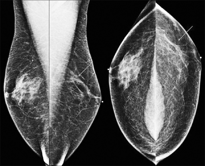
Medial lateral oblique and craniocaudal views of the bilateral breasts show flame-shaped gynecomastia of the right breast. The left breast shows microcalcifcations (arrows) for which magnification views have been performed.
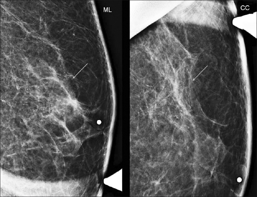
Figure 1
Medial lateral oblique and craniocaudal views of the bilateral breasts show flame-shaped gynecomastia of the right breast. The left breast shows microcalcifcations (arrows) for which magnification views have been performed.
Medial lateral and craniocaudal magnification views of the left breast show suspicious microcalcifications in the retroareolar region extending to the upper outer quadrant (arrows).
Figure 2
Medial lateral and craniocaudal magnification views of the left breast show suspicious microcalcifications in the retroareolar region extending to the upper outer quadrant (arrows).

An ultrasound examination of both breasts was performed utilizing a 12 MHz transducer (Toshiba Alplio MX). The area of pain and retroareolar density in the right breast seen on mammogram corresponded to vague heterogeneous glandular tissue, consistent with gynecomastia, by ultrasound [Figure 3]. No sonographic abnormality was noted in the area of regional microcalcifications in the left breast. Normal left axillary lymph nodes were present.
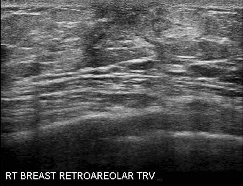
Transverse ultrasound image of the right breast demonstrating retroareolar glandular tissue consistent with gynecomastia. No suspicious masses were identified in the right breast.
Figure 3
Transverse ultrasound image of the right breast demonstrating retroareolar glandular tissue consistent with gynecomastia. No suspicious masses were identified in the right breast.

The patient subsequently underwent stereotactic core biopsy (9 gauge Suros Eviva) of the suspicious microcalcifications in the left breast. Pathology results showed ductal carcinoma in situ grade II, with intermediate nuclear grade features in multiple core fragments [Figure 4]. Estrogen and progesterone receptors were positive. The patient was treated with left total mastectomy and sentinel lymph node biopsy, which was negative. Surgical pathology confirmed DCIS nuclear grade II (intermediate), with all surgical margins uninvolved by DCIS.

Microscopic findings. (a) Ductal carcinoma in situ with intraductal proliferation of small, monomorphic cells, forming secondary microlumina, with clustered microcalcifications (hematoxylin and eosin stain) (4×). (b) Punctate necrosis is present in some involved ducts (hematoxylin and eosin stain) (4×). (c) Microcalcifications are present within the ducts and stroma (hematoxylin and eosin stain) (10×).
Figure 4
Microscopic findings. (a) Ductal carcinoma in situ with intraductal proliferation of small, monomorphic cells, forming secondary microlumina, with clustered microcalcifications (hematoxylin and eosin stain) (4×). (b) Punctate necrosis is present in some involved ducts (hematoxylin and eosin stain) (4×). (c) Microcalcifications are present within the ducts and stroma (hematoxylin and eosin stain) (10×).

DISCUSSION

Most cases of male DCIS present with symptoms on the affected side, such as, palpable mass or bloody nipple discharge.[1] Our case is unusual in that the patient presented with symptoms of gynecomastia in the right breast, and the cancer was an incidental finding in the asymptomatic left breast. The patient was completely asymptomatic on the left with no pain, palpable mass or nipple discharge. Other authors have reported cases of concurrent gynecomastia and DCIS in the same breast, in male patients.[26]

Ductal carcinoma in situ of the male breast is rare, accounting for only about 7% of all male breast cancer cases.[1] Of all patients (male and female) with DCIS, 30 - 50% develop invasive cancer in the following 10 - 20 years, so the actual prevalence may be higher.[2] A vast majority of male breast cancers are ductal, and usually present late, with approximately 40% having stage III or IV disease.[4] Men are less likely than women to be diagnosed in the early stages, but diagnosis at the non-invasive stage has increased. This increase in early diagnosis may be due to improved awareness of male breast cancer. Factors that increase the risk of male breast cancer include high estrogen states, Klinefelter's syndrome, family history, BRCA 2 gene, hyperprotactinemia, and prior radiation.[78] No increased breast cancer risk associated with gynecomastia in males has been found.[24]

The exact cause of DCIS in males in unknown, as they lack the terminal duct lobular units where DCIS often originates in women. It is hypothesized, however, that DCIS originates in the duct epithelium in men.[2] Mammography for male breast cancer has a reported sensitivity of 92% and specificity of 91%.[9] Calcifications are less frequently associated with male breast cancer than female breast cancer. Malignant calcifications in the male breast often have a more benign appearance and can be more scattered or round.[910]

Management of male breast cancer is largely based on studies that deal with female breast cancer. Typical treatment for males with DCIS is total mastectomy without axillary node dissection.[18] In a series of 31 cases of pure DCIS in men by Cutuli et al., 19 cases underwent axillary node dissection, with all sampled lymph nodes negative.[3] Recurrence of male DCIS following total mastectomy has yet to be described.[2] In the same series of 31 cases of male DCIS, Cutuli et al., described only four recurrences, none of which were in patients treated with total mastectomy.[3] No further adjuvant therapy is necessary, and prognosis for DCIS in males is excellent.

Non-invasive breast cancers in men are symptomatic at presentation. For men, there is no standardized routine screening program for breast cancer. Our case emphasizes that the contralateral breast should also be imaged in males presenting with unilateral breast symptoms.

Source of Support: Nil

Conflict of Interest: None declared.

REFERENCES

  1. , , , , , , . Ductal carcinoma in situ of the male breast. Cancer. 1994;74:1289-93.
    [Google Scholar]
  2. , , . Ductal carcinoma in situ in a 25-year-old man presenting with apparent unilateral gynecomastia. Curr Oncol. 2010;17:133-7.
    [Google Scholar]
  3. , , , , , , . Ductal carcinoma in situ of the male breast.Analysis of 31 cases. Eur J Cancer. 1997;33:35-8.
    [Google Scholar]
  4. , , , . Male breast cancer. Lancet. 2006;367:595-604.
    [Google Scholar]
  5. , , . Ductal carcinoma in situ and bilateral atypical ductal hyperplasia in a 23-year-old man with gynecomastia. Am Surg. 2011;77:1272-3.
    [Google Scholar]
  6. , , , , . Ductal carcinoma in situ of the male breast presenting as adolescent unilateral gynaecomastia. J Plast Reconstr Aesthet Surg. 2011;64:1684-6.
    [Google Scholar]
  7. , , . Gynaecomastia and breast cancer in men. BMJ. 2008;336:709-13.
    [Google Scholar]
  8. , , , , . Synchronous bilateral noninvasive ductal carcinoma of the male breast: A case report. Breast Cancer. 2003;10:163-6.
    [Google Scholar]
  9. , , , , , , . The diagnostic accuracy of mammography in the evaluation of male breast disease. Am J Surg. 2001;181:96-100.
    [Google Scholar]
  10. , , , . Imaging male breast cancer. Clin Radiol. 2011;66:1079-85.
    [Google Scholar]

Fulltext Views
10,329

PDF downloads
2,236
View/Download PDF
Download Citations
BibTeX
RIS
Show Sections