
Translate this page into:
Apparent Diffusion Coefficient Measurement in Mediastinal Lymphadenopathies: Differentiation between Benign and Malignant Lesions
Address for correspondence: Dr. Deniz Alis, Department of Radiology, Cerrahpasa Faculty of Medicine, Istanbul University, Fatih, Istanbul 34098, Turkey. E-mail: drdenizalis@gmail.com
-
Received: ,
Accepted: ,
This is an open access article distributed under the terms of the Creative Commons Attribution-NonCommercial-ShareAlike 3.0 License, which allows others to remix, tweak, and build upon the work non-commercially, as long as the author is credited and the new creations are licensed under the identical terms.
This article was originally published by Medknow Publications & Media Pvt Ltd and was migrated to Scientific Scholar after the change of Publisher.
Abstract
Objectives:
We aimed to prospectively assess the diagnostic value of apparent diffusion coefficient (ADC) measurement in the differentiation of benign and malignant mediastinal lymphadenopathies.
Materials and Methods:
The study included 63 consecutive patients (28 women, 35 men; mean age 59.3 years) with 125 mediastinal lymphadenopathies. Echoplanar diffusion-weighted magnetic resonance imaging of the mediastinum was performed with b-factors of 0 and 600 mm2/s before mediastinoscopy and mediastinotomy, and ADC values were measured. The ADC values were compared with the histological results, and statistical analysis was done. P < 0.05 was considered statistically significant.
Results:
The mean ADC value of malignant mediastinal lymphadenopathy (1.030 ± 0.245 × 10⁻³ mm2/s) was significantly lower (P < 0.05) when compared to benign lymphadenopathies (1.571 ± 0.559 × 10⁻³ mm2/s). For differentiating malignant from benign mediastinal lymphadenopathy, the best result was obtained when an ADC value of 1.334 × 10⁻³ mm2/s was used as a threshold value; area under the curve 0.848, accuracy 78.4%, sensitivity 66%, specificity of 86%, positive predictive value 76.7%, and negative predictive value of 79.2%. Interobserver agreement was excellent for ADC measurements.
Conclusions:
ADC measurements could be considered an important supportive method in differentiating benign from malignant mediastinal lymphadenopathies.
Keywords
Apparent diffusion coefficient
diffusion
lymph node
mediastinum
magnetic resonance imaging


Introduction
Mediastinal lymphadenopathies are frequently seen with a neoplastic or an inflammatory disease in which knowledge of lymph node involvement is a significant prognostic factor in lung malignancy. Therefore, detection and characterization of mediastinal lymphadenopathies is a keystone for staging and treatment of the patients with lung cancer.[12] Even with the help of new technology such as computed tomography (CT), magnetic resonance imaging (MRI), 18-fluoro-2-deoxy-glucose positron emission tomography (PET), bronchoscopy, mediastinoscopy, and thoracoscopy, it is still difficult to clarify whether lung cancer metastasis or inflammatory diseases are the cause of the enlarged mediastinal lymph nodes.[3]
CT scanning has been used as powerful tool for the presence and dimension of mediastinal lymph nodes. Although CT provides valuable information for detection of enlarged lymph nodes, it could not accurately differentiate malignant from inflammatory nodes.[45] CT sensitivity and specificity are up to 60%, which are not adequate for clinical decision-making.[67]
PET-CT scan is sensitive enough to detect suspicious lymph nodes, false positive results of mediastinal, and hilar lymph nodes were reported to be due to pneumoconiosis, silicosis, pulmonary tuberculosis, and sarcoidosis.[8]
Mediastinoscopy or CT-guided biopsy is gold standard for diagnosis. Main disadvantages of these approaches are inadequate tissue sampling and possible complications.[9]
Conventional MRI (contrast enhanced and noncontrast enhanced T1-weighted and T2-weighted axial) is able to determine the presence and size of nodes; however, it cannot be used to reliably differentiate benign from malignant mediastinal lymph nodes.[10] Diffusion-weighted imaging (DWI) is a magnetic resonance technique based on the Brownian motion of water to calculate diffusion of water protons through tissue. The specific diffusion capacity of a biologic tissue is referred to as apparent diffusion coefficient (ADC). ADC measurements have proven valuable for the detection and characterization of tumors in such parenchymal organs as the liver, kidney, pancreas, bile duct, gallbladder, and prostate, in recent years.[11121314] The purpose of this study was to assess the diagnostic value of the ADC measurements in the differentiation of benign and malignant mediastinal lymphadenopathies.
Materials and Methods
The Local Research Ethics Committee approved this prospective study, and written informed consent was obtained from each patient.
Patients
Sixty-three patients (28 women, 35 men; mean age, 59.3 years; age range, 25–74 years) with mediastinal lymphadenopathies detected by chest CT were referred for MRI of the mediastinum between 2013 and 2015. We excluded patients who had been performed mediastinoscopy, mediastinotomy, chemotherapy, or radiotherapy after detection of mediastinal lymph nodes by chest CT.
Magnetic resonance technique
Sixty-three patients with 125 mediastinal lymph nodes underwent MRI examinations using a 1.5 Tesla MRI unit (Avanto, Siemens Medical Systems, Enlargen, Germany).
All MRI examinations were performed with a high-performance gradient subsystem (maximum gradient strength of 45 mT/m) using a body phased-array coil.
Patients were laid supine position and instructed not to move during the examination.
The DW-MRI parameters were as follows: TR/TE = 3200/75 ms; slice thickness = 5 mm; interslice gap = 0.5 mm; number of slices = 35; matrix size = 153 × 192, with reconstruction to 256 × 256; field of view = 325 mm × 325 mm; bandwidth = 1736 hz/pixel; number of signal averages = 2.
Image analysis
Quantitative measurements of DWI were interpreted by two radiologists on a picture archiving and communication system viewing station. Both of the radiologists were blinded to the patients’ clinical findings and the results of the prior imaging studies. First reader (FEU) was a 4th-year radiology resident, and second reader (DCO) was a radiologist with an experience of 8 years in thoracic MRI at the time of data analysis.
The first reader evaluated ADCs twice in a 1-week period; he traced the same procedure to assess intraobserver reproducibility, and his first measurement was compared with the measurement obtained by the second reader to assess interobserver agreement.
The term “mediastinal lymphadenopathy” describes a lymph node, in which defined as more than 1 cm or larger in short-axis diameter.[15] In our study, we accepted lymph nodes as a lymphadenopathy according to this definition. To determine the location, size, and the presence of cystic-necrotic parts of the lymph nodes, each lesion was interpreted on T2-weighted images initially. Quantitative ADC maps were calculated on voxel-by-voxel basis using commercial workstation (Syngo Via, Siemens Medical Healthcare, Germany) for combination of b-values 0 and 600 mm2/s. The region of interests (ROIs) was placed on the lymph nodes and size of the ROIs kept as large as possible. We avoided surrounding lung tissue, cystic areas, and necrotic parts. Each observer performed total 4–6 ADC measurements in several slices from the lymph node and the average of the mean ADC values were calculated [Figures 1 and 2].
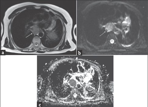
- A 76-year-old woman with known lung carcinoma. Magnetic resonance imaging of the mediastinum. (a) Axial T2-weighted image shows an enlarged subcarinal lymphadenopathy (arrow). (b) On axial diffusion-weighted magnetic resonance image at b = 600 s/mm2, the lymphadenopathy is hyperintense (arrow). (c) Region of interest was placed on hypointense lymphadenopathy (arrow) with an apparent diffusion coefficient value = 0.97 ± 0.23 × 10⁻³ mm2/s.
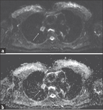
- A 48-year-old woman with known sarcoidosis. Magnetic resonance imaging of the mediastinum. (a) On axial diffusion-weighted magnetic resonance image at b = 600 s/mm2, the lymphadenopathy is hypointense (arrow). (b) The region of interest was placed on slightly hyperintense lymphadenopathy (arrow) with an apparent diffusion coefficient value = 1.63 ± 0.1 × 10⁻³ mm2/s.
Lymph node sampling and histopathologic examination
Sixty-three patients underwent mediastinoscopy or mediastinotomy. As a consequence of this, final diagnosis of lymph nodes was established based on the histologic examination. The histopathology reports were retrospectively reviewed.
One hundred and twenty-five lymph nodes were analyzed histopathologically. The ADC values were compared with pathologic results on a lesion-by-lesion basis.
Statistical analysis
Statistical analyses were performed using SPSS software (version 16.0; SPSS Inc., Chicago, IL, USA). The Kolmogorov–Smirnov test showed that the data were not normally distributed, so the differences in ADCs were analyzed using the Mann–Whitney U-test.
P < 0.05 was considered statistically significant. Receiver operating characteristic (ROC) analysis was performed to evaluate the diagnostic performance of the mean ADC measurements in differentiating malignant from benign lesions and to describe the sensitivity and specificity. An optimum cut-off point was hence determined as the value that best discriminates between the two groups in terms of maximum sensitivity and a minimum number of false positive results.
Inter-reader agreement for benign and malignant lymph nodes in the quantitative analysis was calculated using intraclass correlation coefficients (ICCs) from a one-way random effects model analysis of variance with the subject as the random effect. A 95% confidence interval was constructed for each ICC. An ICC >0.80 indicated excellent agreement.
Results
Localization of the lymph nodes was determined according to Union Internationale Contre le Cancer.[16] Lymph nodes in our study were including upper paratracheal (30), lower paratracheal (43), 27 subcarinal (27), hilar (16), prevertebral (6), and paraaortic (3). Dimension (short axis) of the lymph nodes was between 0.6 cm and 5.5 cm (median dimension = 2.26 cm).
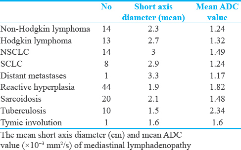
A total of 125 lymph nodes were observed in 65 patients. The histopathological diagnosis of the mediastinal malignant lymphadenopathy was nonsmall cell lung cancer (SCLC) (n = 14), SCLC (n = 8), non-Hodgkin lymphoma (n = 14), and Hodgkin lymphoma (n = 13). Mean ADC values and mean short-axis diameter of these groups were summarized in Table 1.
There was 1 metastatic lymph node from distant site primary tumor (colorectal carcinoma).
The histopathological diagnosis of the mediastinal benign lymphadenopathy was reactive lymphoid hyperplasia in 44 patients, sarcoidosis in 20 patients, tuberculous nodes in 10 patients, and thymic involution in 1 patient. The dimension (short axis) of mediastinal lymph nodes varied from 0.6 to 5.5 cm.
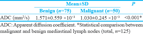
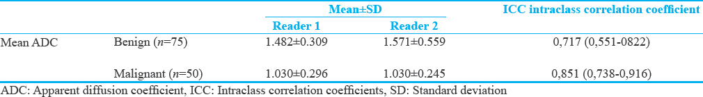
The mean ADC value of malignant mediastinal lymphadenopathy was 1.030 ± 0.245 × 10⁻³ mm2/s and that of benign lymphadenopathy was 1.571 ± 0.559 × 10⁻³ mm2/s. There was a significant difference in the ADC values between malignant and benign lymph nodes (P < 0.001). The mean and standard deviations of malignant and benign mediastinal lymphadenopathy are shown in Table 2.
The receiver operating characteristic curves for the differentiation of benign and malignant lymph node based on ADC values are shown in Figure 2.
In our study, the ADC cutoff value for differentiating malignant from benign nodes that maximize the accuracy was 1.334 × 10⁻³ mm2/s and its sensitivity, specificity, positive predictive value, negative predictive value, and accuracy were 66%, 86%. 76.7%, 79.2% 78.4%, respectively. The area under the curve was 0.848 [Figure 3].
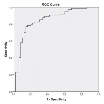
- Receiver operating characteristic curve for the differentiation of benign and malign mediastinal lymph nodes based on apparent diffusion coefficient measurements.
The result of the interobserver variability is shown in Table 3. Interobserver agreement was excellent for both benign (ICC: 0.717) and malignant (ICC 0.851) lymph nodes groups.
Discussion
In our study, we found that the mean ADC value of metastatic mediastinal lymph nodes (1.030 ± 0.245 × 10⁻³ mm2/s) was significantly lower than that of benign lymph nodes (1.571 ± 0.559 × 10⁻³ mm2/s).
Mediastinal lymph nodes may be enlarged for a variety of inflammatory, infectious, or malignant reasons. Differentiation of mediastinal lymph node is essential not only for diagnosis and staging of malignant diseases but also for determining treatment and follow-up.[171819]
DW-MRI is a noninvasive technique that measures the motion of water in the extracellular space. Metastatic lymph nodes have a reduced diffusivity occurred due to hypercellularity, or increased nuclear-to-cytoplasmic ratio.[20] The specific diffusion capacity of a biologic tissue is referred to as ADC.[21] The restricted ADC values in metastases may be related with the increased cell density and the ratio of nucleus to cytoplasm.[2223]
The previous studies with DW-MRI successfully showed the metastatic nodes, benign lymphadenopathy, and nodal lymphomas in the head and neck.[2425] Wang et al. performed DWI to head or neck lesions and found that the mean ADC value of malignant lesions (1.13 ± 0.43 × 10⁻³ mm2/s) was less than that of benign solid masses (1.560 ± 0.51 × 10⁻³ mm2/s).[20] Kosucu et al. and Abdel Razek et al. reported a significant difference in the ADC value between benign and metastatic mediastinal lymph nodes (P < 0.005 and P = 0.001).[2627]
Conversely, Sumi et al. found that the ADC was significantly greater in metastatic lymph nodes (0.410 ± 0.105 × 10⁻³ mm2/s, P < 0.01) than in benign lymphadenopathy (0.302 ± 0.062 × 10⁻³ mm2/s) in cervical lymph nodes.[24] The cause of this contradiction may be attributable to the placement of the ROI, which included necrotic areas.
Our study had several limitations. First, histopathological types of mediastinal lymph nodes demonstrated heterogeneous distribution. Future studies are recommended to evaluate each specific pathological entity. Second, small lymph nodes (<0.6 cm) were excluded from the evaluation to obtain reliable ADC values. Third, we did not compare DW-MRI with PET-CT. Fourth, we did not take into account the shape and intensity of the mediastinal lymph nodes.
Conclusion
Our study demonstrated that the mean ADC value of the metastatic lymph nodes was significantly lower than that of the nonmetastatic lymph nodes in patients with mediastinal lymph nodes. We suggested that reduced ADC values of metastatic lymph nodes compared to nonmetastatic lymph nodes might help the diagnosis.
Financial support and sponsorship
Nil.
Conflicts of interest
There are no conflicts of interest.
Available FREE in open access from: http://www.clinicalimagingscience.org/text.asp?2017/7/1/12/201644
References
- Imaging of mediastinal lymph nodes: CT, MR, and FDG PET. Radiographics. 1998;18:1061-9.
- [Google Scholar]
- MR imaging of thoracic lymph nodes. A comparison of computed tomography and positron emission tomography. Magn Reson Imaging Clin N Am. 2000;8:33-41.
- [Google Scholar]
- The clinical application value of PET/CT in adenocarcinoma with bronchioloalveolar carcinoma features. Ann Nucl Med. 2010;24:541-7.
- [Google Scholar]
- T1 lung cancer: Prevalence of mediastinal nodal metastases and diagnostic accuracy of CT. Radiology. 1993;186:129-32.
- [Google Scholar]
- Is it possible to differentiate malignant mediastinal nodes from benign nodes by size? Reevaluation by CT, transesophageal echocardiography, and nodal specimen. Chest. 1996;110:1004-8.
- [Google Scholar]
- The mediastinum in non-small cell lung cancer: CT-surgical correlation. AJR Am J Roentgenol. 1984;142:1101-5.
- [Google Scholar]
- Imaging of the pulmonary hilum: A prospective comparative study in patients with lung cancer. AJR Am J Roentgenol. 1985;145:245-8.
- [Google Scholar]
- Mediastinal lymphadenopathy: Diagnostic yield of transbronchial mediastinal lymph node biopsy with CT fluoroscopic guidance-initial experience. Radiology. 2000;216:764-7.
- [Google Scholar]
- Differentiation of metastatic versus non-metastatic mediastinal lymph nodes in patients with non-small cell lung cancer using respiratory-triggered short inversion time inversion recovery (STIR) turbo spin-echo MR imaging. Eur J Radiol. 2002;44:216-24.
- [Google Scholar]
- Role of diffusion-weighted magnetic resonance imaging in the diagnosis of gallbladder cancer. J Magn Reson Imaging. 2013;38:127-37.
- [Google Scholar]
- Segmental liver hyperintensity in malignant biliary obstruction on diffusion weighted MR imaging: Associated MRI findings and relationship with serum alanine aminotransferase levels. Br J Radiol. 2012;85:22-8.
- [Google Scholar]
- Prostate MRI: Diffusion-weighted imaging at 1.5T correlates better with prostatectomy Gleason grades than TRUS-guided biopsies in peripheral zone tumours. Eur Radiol. 2012;22:468-75.
- [Google Scholar]
- New MR imaging criteria with a diffusion-weighted sequence for the diagnosis of hepatocellular carcinoma in chronic liver diseases. J Hepatol. 2011;55:126-32.
- [Google Scholar]
- Agreement of mediastinal lymph node size between computed tomography and endobronchial ultrasonography: A study of 617 patients. Ann Thorac Surg. 2015;99:1894-8.
- [Google Scholar]
- CT demonstration of the 1996 AJCC-UICC regional lymph node classification for lung cancer staging. Radiographics. 1999;19:899-900.
- [Google Scholar]
- Prospective evaluation of mediastinoscopy for assessment of carcinoma of the lung. J Thorac Cardiovasc Surg. 1986;91:53-6.
- [Google Scholar]
- Head and neck lesions: Characterization with diffusion-weighted echo-planar MR imaging. Radiology. 2001;220:621-30.
- [Google Scholar]
- Lung carcinoma: Diffusion-weighted mr imaging – Preliminary evaluation with apparent diffusion coefficient. Radiology. 2007;243:570-7.
- [Google Scholar]
- Relationship between bone marrow cellularity and apparent diffusion coefficient. J Magn Reson Imaging. 2001;13:757-60.
- [Google Scholar]
- Discrimination of axillary metastatic from nonmetastatic lymph nodes with PROPELLER diffusion-weighted MR imaging in a metastatic breast cancer model and its correlation with cellularity. J Magn Reson Imaging. 2012;36:624-31.
- [Google Scholar]
- Discrimination of metastatic cervical lymph nodes with diffusion-weighted MR imaging in patients with head and neck cancer. AJNR Am J Neuroradiol. 2003;24:1627-34.
- [Google Scholar]
- MR microimaging of benign and malignant nodes in the neck. AJR Am J Roentgenol. 2006;186:749-57.
- [Google Scholar]
- Mediastinal lymph nodes: Assessment with diffusion-weighted MR imaging. J Magn Reson Imaging. 2009;30:292-7.
- [Google Scholar]
- Characterization of mediastinal lymphadenopathy with diffusion-weighted imaging. Magn Reson Imaging. 2011;29:167-72.
- [Google Scholar]


