Translate this page into:
Acquired Arteriovenous Fistula of the Breast Following Ultrasound Guided Biopsy of Invasive Ductal Carcinoma
Address for correspondence: Dr. Adam Gregg, Department of Radiology and Radiological Science, Medical University of South Carolina, 96 Jonathan Lucas Street, MSC 323 Charleston, SC 29425, USA. E-mail: greggat@musc.edu
-
Received: ,
Accepted: ,
This is an open-access article distributed under the terms of the Creative Commons Attribution License, which permits unrestricted use, distribution, and reproduction in any medium, provided the original author and source are credited.
This article was originally published by Medknow Publications & Media Pvt Ltd and was migrated to Scientific Scholar after the change of Publisher.
Abstract
Image guided large-core breast biopsies are commonly performed procedures with relatively rare complications. The majority of these complications are minor, though at times more significant vascular injuries can occur with these biopsies as demonstrated by this case. Patient developed a pulsatile vascular breast mass after an ultrasound guided breast biopsy of invasive ductal carcinoma. Sonographic evaluation of this new breast mass demonstrated this mass to represent an arteriovenous fistula (AVF). Though multiple therapies are available for an iatrogenic fistula within the breast, the AVF was surgically excised in this case as it was immediately adjacent to a known cancer.
Keywords
Arteriovenous fistula
biopsy
breast
ultrasound
INTRODUCTION

Large-core breast biopsy is a widely used tool, with increasing acceptance of vacuum-assisted large-core breast biopsies.[1] Reported complication after image guided needle core biopsy of the breast is rare, with the most common complications representing hematoma, bleeding, pain and infection.[12] The majority of these complications are minor and clinically insignificant.[2] However, significant vascular injuries in the breast have been reported, including post-procedural pseudoaneurysms and arteriovenous fistulas (AVFs).[345]
CASE REPORT
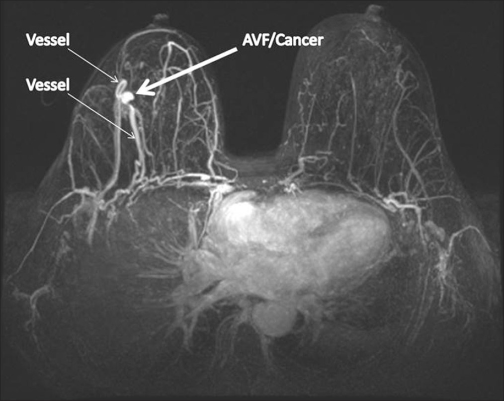
A 58-year-old female presented for her annual screening mammogram. At that time, a new mass was identified in the right breast at 9 o’clock, middle depth region. Targeted ultrasound showed a 1.7 cm × 1.4 cm × 1.3 cm irregular hypoechoic mass to correlate with the mammographic abnormality. Ultrasound guided biopsy of the right breast mass was subsequently performed with a 14 gauge vacuum assisted biopsy device. At the time of biopsy, a significant amount of bleeding was noted after the final core biopsy. Bleeding was controlled with manual compression. A marker clip was placed. Post-procedure mammograms demonstrated a large hematoma at the biopsy site with the marker clip associated with the mass. Pathology results showed this mass represented invasive ductal carcinoma. Subsequent magnetic resonance imaging (MRI) demonstrated a new round enhancing mass with rapid wash-in and rapid wash-out characteristics. The mass measuring 0.8 cm × 0.8 cm was seen at the site of biopsy adjacent to the enhancing known cancer that measured up to 2.1 cm [Figure 1a]. Large vessels were seen coursing to and from the new arterially enhancing mass [Figure 1a]. Clinical examination revealed a new pulsatile mass within the right breast.

- 58-year-old female diagnosed with arteriovenous fistula of the right breast with adjacent invasive ductal carcinoma. Axial maximum intensity projections magnetic resonance image demonstrates an arterially enhancing mass (thick arrow) with prominent feeding and draining vessels (thin arrows).
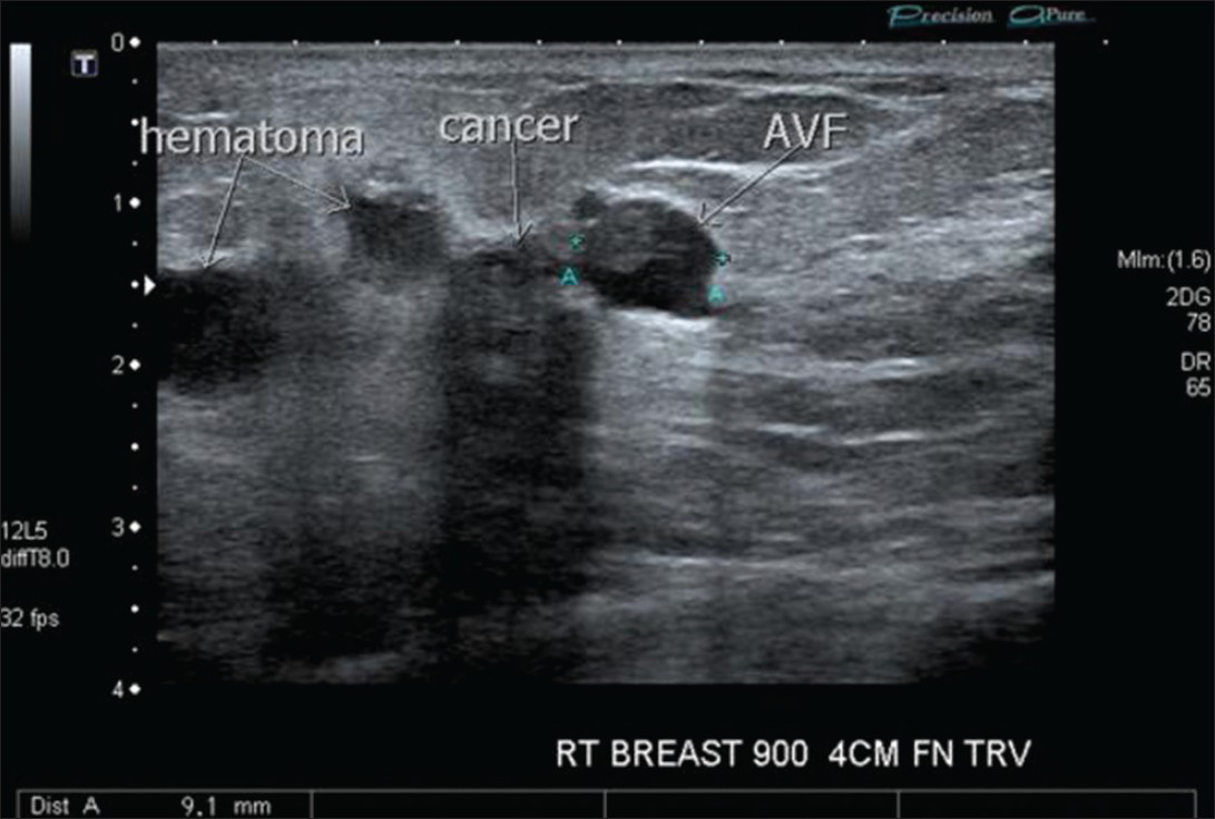
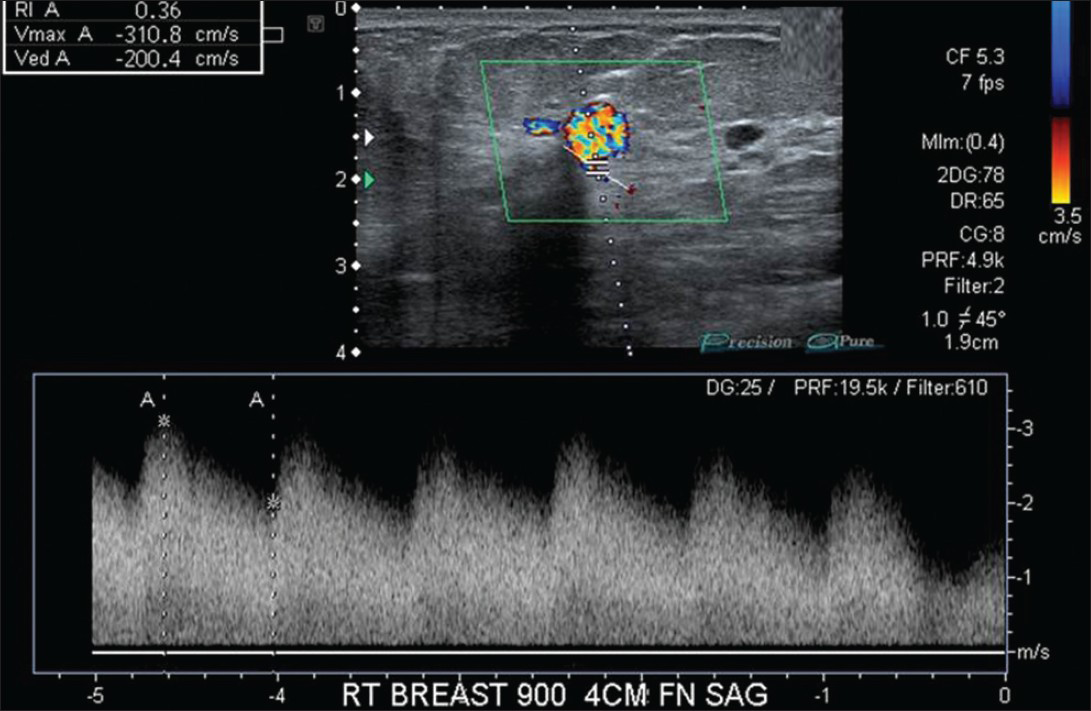
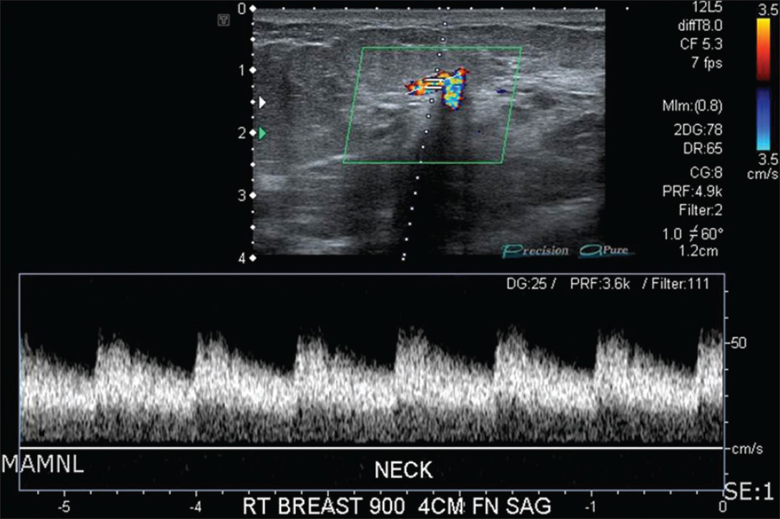
Ultrasound examination of the breast was performed to evaluate the new suspected vascular mass. A vascular mass was seen adjacent to the known carcinoma within a region of hematoma and measured 0.9 cm × 0.8 cm × 0.6 cm [Figure 1b]. Two large vessels were seen in continuity with this vascular mass. A high velocity flow, low resistance Doppler wave form was seen in the mass consistent with an AVF [Figure 1c]. Arterialization of the draining vein was also noted [Figure 1d]. The vascular mass and adjacent known neoplasm were excised using intraoperative ultrasound. The pathology demonstrated invasive ductal carcinoma adjacent to a vascular lesion.

- 58-year-old female diagnosed with arteriovenous fistula (AVF) of the right breast with adjacent invasive ductal carcinoma. Transverse sonogram shows an AVF with internal thrombus adjacent to patient's known hypoechoic and shadowing invasive ductal carcinoma with surrounding hypoechoic hematoma.

- 58-year-old female diagnosed with arteriovenous fistula (AVF) of the right breast with adjacent invasive ductal carcinoma. Sagittal color Doppler sonogram with spectral waveform imaging shows high velocity, low resistance wave forms within the AVF.

- 58-year-old female diagnosed with arteriovenous fistula of the right breast with adjacent invasive ductal carcinoma. Sagittal color Doppler sonogram with spectral waveform imaging shows arterialization of the draining vein.
DISCUSSION
An AVF is an abnormal communication between an artery and a vein, bypassing an interposed capillary system with shunting of blood.[6] AVFs are classified as being congenital or acquired. Congenital AVFs (arteriovenous malformations) typically occur as a result of failure of differentiation between an artery and vein during embryology.[3] Acquired AVFs can be the result of trauma, neoplasm, erosion of an arterial aneurysm or surgically created hemodialysis fistulas.[6] Penetrating trauma has been reported as the most common cause of acquired traumatic AVFs, accounting for 3% of all vascular injuries. Acquired AVFs generally demonstrate one large feeding artery and one large draining vein. Congenital AVFs on the other hand typically demonstrate multiple feeding arteries with multiple venous aneurysms.[3]
AVFs may remain clinically silent but can also present with pulsatile hematomas, palpable thrills or audible bruits.[3] Repair of the fistula is important as the fistula can enlarge increasing the risk of rupture and embolization.[4] Ultrasound is a good non-invasive imaging modality to initially investigate suspected AVFs while angiography remains the gold standard.[3] Low resistance flow in the supplying artery, high-velocity arterialized waveform in the draining vein and turbulent high-velocity flow spectrum at the junction of the artery and vein are considered the criteria for the diagnosis of AVF by color Doppler ultrasound and spectral waveform analysis. Computed tomography and MRI angiography demonstrate early contrast filling in the vein during the arterial phase. Angiography is most helpful for mapping the feeding arteries and draining veins for possible endovascular therapy, to include catheter embolization.[6] Surgical ligation was the treatment of choice for the prior two cases of AVFs, though surgical excision using sonographic guidance was opted for in this case as it was removed en-bloc with the adjacent cancer.[34]
CONCLUSION
Although complications related to image guided biopsies in the breast are relatively rare, they can and do occur. Vascular injuries, particularly pseudoaneurysms and AVFs, are rare complications that need to be considered to assure appropriate treatment. Ultrasound is a suitable imaging modality to evaluate a suspected AVF to help guide appropriate therapy as in this case where it was used to diagnose as well as guide treatment.
Available FREE in open access from: http://www.clinicalimagingscience.org/text.asp?2013/3/1/38/119019
Source of Support: Nil
Conflict of Interest: None declared.
REFERENCES
- Vacuum-assisted large-core breast biopsy: Complications and their incidence. Can Assoc Radiol J. 2000;51:232-6.
- [Google Scholar]
- Pseudoaneurysm of the breast: Case study and review of literature. Br J Radiol. 2004;77:694-7.
- [Google Scholar]
- Case report of pseudoaneurysm caused by core needle biopsy of the breast. Breast Cancer. 2010;17:75-8.
- [Google Scholar]


