Translate this page into:
A Case of Mitral Valve Tophus in a Patient with Severe Gout Tophaceous Arthritis
Address for correspondence: Dr. Atooshe Rohani, Cardiac Research Center, School of Medicine, Mashhad University of Medical Sciences, Mashhad, Iran. E-mail: rohania@mums.ac.ir
-
Received: ,
Accepted: ,
This is an open-access article distributed under the terms of the Creative Commons Attribution License, which permits unrestricted use, distribution, and reproduction in any medium, provided the original author and source are credited.
This article was originally published by Medknow Publications & Media Pvt Ltd and was migrated to Scientific Scholar after the change of Publisher.
Abstract
A few cases of cardiac valve tophi have been reported in literature. In this case report, the echocardiographic characteristics of the hyperechoic mass in the posterior leaflet mitral valve, intact mitral valve ring, and the occurrence of severe tophaceous gout arthritis suggested the diagnosis of a gout tophus on the mitral valve.
Keywords
Arthritis
echocardiogram
gout
mitral valve
tophus
INTRODUCTION

Gout usually presents with recurrent episodes of joint inflammation, which over time lead to tophus formation and joint destruction. There is a strong association of gout and cardiovascular disease, but one does not directly cause the other.[1]
We present a case of a patient suffering from gout arthritis with tophous on the mitral valve that was detected with echocardiography.
CASE REPORT
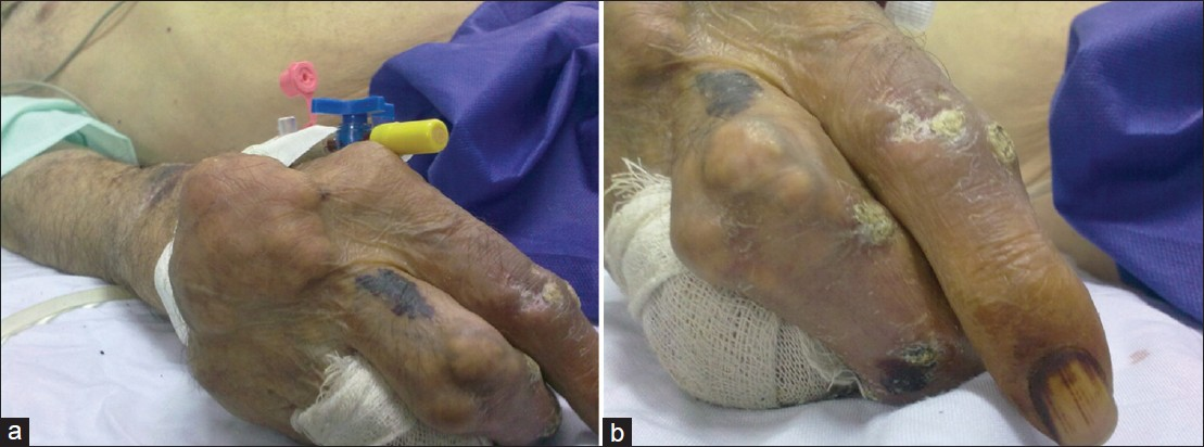
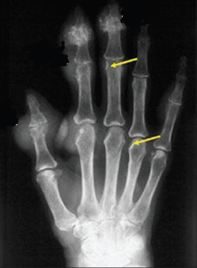
A 75-year-old man presented with multiple tophi over bilateral hands, feet, elbows, knees, and ankles that had worsened over 10 years. He was admitted to the Internal Medicine department due to an acute attack of gouty arthritis. Since gout is associated with increased risk of congestive heart failure and systolic dysfunction,[1] he was referred to the Cardiology department for echocardiography. Except for gout arthritis, the patient's history was unremarkable. He was not on any medications. Dermatological examination revealed multiple, mobile, skin-to-yellowish colored, firm, dermal and subcutaneous nodules and large globose tumors, located on dorsa of the hands overlying inter-phalangeal joints, wrists, ankles, and interphalangeal joints of the feet. Some lesions had ulcerated discharging white chalky material [Figures 1a and b]. The associated joints were deformed. Hematological and biochemical examinations revealed hemoglobin level of 14 gm/dl, raised serum uric acid (11.4 mg/dl, normal: 2-7.4 mg/dl), blood urea (110 mg/ dl, normal: 10-50 mg/dl), and serum creatinine (1.8.0 mg/dl, normal: 0.5-1.3 mg/dl). Liver function test, urinalysis, and serum electrolytes were within normal limits. Rheumatoid factor was negative. Radiographic evaluation of both hands showed soft-tissue swelling and periarticular erosions in the lower end of radius and interphalangeal joints with sclerotic margins and overhanging edges [Figure 2].

- (a and b) Multiple nodules overlying deformed joints seen in the hand of a chronic patient with tophaceous gout.

- Soft tissue radiograph of hand shows swelling and arrows point to bone erosion.
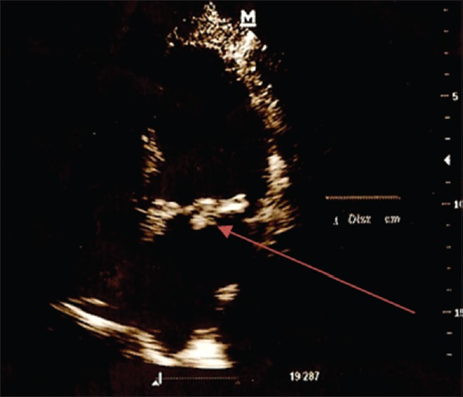
Transthoracic echocardiogram showed a hyperechoic, oval-shaped mass measuring 1.5 × 1 ×1 cm on posterior mitral leaflet not reducing significantly the opening area of the valve (3.0 cm2), but he had trivial mitral regurgitation [Figure 3]. Left atrium in M-mode was 36 mm and left ventricle diameters (systolic 25 mm and diastolic 38 mm) were normal. Mild aortic regurgitation was present, but tricuspid and pulmonary valves were unaffected and functioning well. Transesophageal echocardiogram confirmed the finding of an isolated mass on posterior mitral leaflet without significant valve dysfunction.

- Transthoracic echocardiogram in apical 4 chamber view: Arrow points to a hyperechoic, oval-shaped mass on the mitral valve.
DISCUSSION
Gout is characterized by recurrent attacks of acute inflammatory arthritis and abnormal metabolism of uric acid, resulting in an excess of uric acid in the tissues and blood. Hyperuricemia is necessary but not sufficient for development of gout. It is definitively diagnosed by detecting uric acid (monosodium urate) crystals in an aspirated sample of the joint fluid. Hard deposits of crystals, tophi, may develop under the skin around the joints. The most important differential diagnosis in gout is septic arthritis. Other differential diagnoses are pseudogout and rheumatoid arthritis. Gouty tophi, in particular when not located in a joint, can be mistaken for basal cell carcinoma. Visceral tophi mainly occur in the urinary tract, such as the kidney, ureter, collectively referred to as urinary tract stones. Gout stones can also be found in the digestive tract. According to current knowledge, the only tissue without the formation of tophi is the brain tissue. Elevated serum urate levels have been shown to correlate with high blood pressure in adults and raises a significant independent risk of death due to cardiovascular disease. Gouty arthritis is reported with an excess risk of acute myocardial infarction, and it is not explained by its well-known risk factors. For those with gout, the risk for CVD mortality was about 30% higher than for those without gout, even after adjustment for expected risk factors.[2]
Very few cases of cardiac valve tophi have been reported.[3] Gout tophi commonly occur in soft tissue and are rare in cardiac valves.[3–7]
In this case report, the echocardiographic characteristics of the hyperechoic mass and the occurrence of severe tophaceous gout artrithis provide a presumptive diagnosis of a tophus on the mitral valve. The exact diagnosis cannot be made without proper histological examination, nonetheless, the patient did not suffer from other clinical features suggestive of infective endocarditis and had no cardiovascular risk factors for coronary artery disease (which is associated with mitral valve annulus calcification). As a result, the occurrence of the hyperechoic mass at the mitral valve was most likely caused by gout.
CONCLUSION
This case suggests when the presence of a hyperechoic lesion on the mitral valve is seen on the echocardiogram, gout arthritis should be considered in the differential diagnosis.
Available FREE in open access from: http://www.clinicalimagingscience.org/text.asp?2012/2/1/68/103058
Source of Support: Nil
Conflict of Interest: None declared.
REFERENCES
- Gout and the risk for incident heart failure and systolic dysfunction. BMJ Open. 2012;2:e000282.
- [Google Scholar]
- Gout and the risk of acute myocardial infarction. Arthritis Rheum. 2006;54:2688-96.
- [Google Scholar]
- A rare and asymptomatic case of mitral valve tophus associated with severe gouty tophaceous arthritis. J Endocrinol Invest. 2004;27:965-6.
- [Google Scholar]
- Gout, just a nasty event or a cardiovascular signal.A study from primary care? Fam Pract. 2003;20:413-6.
- [Google Scholar]


