More results...

Generic selectors
Exact matches only
Search in title
Search in content
Post Type Selectors
Filter by Categories
Abdominal Radiology, Original Research
Abdominal Radiology, Pictorial Essay
Anthropology and Dental Radiology, Original Research
Breast Imaging, Case Report
Breast Imaging, Original Research
Breast Imaging, Pictorial Essay
Breast Imaging, Review Article
Cardiopulmonary Imaging, Case Report
Cardiopulmonary Imaging, Case Series
Cardiopulmonary Imaging, Education
Cardiopulmonary Imaging, Original Research
Cardiopulmonary Imaging, Pictorial Essay
Cardiopulmonary Imaging, Review Article
Case Report
Case Report, Breast Imaging
Case Report, Cardiopulmonary Imaging
Case Report, Diagnostic Radiology
Case Report, Gastrointestinal Imaging
Case Report, Genitourinary and Gynecologic Imaging
Case Report, Neuroradiology/Head and Neck Imaging
Case Report, Vascular and Interventional Radiology
Case Series
Case Series, Abdominal Radiology
Case Series, Diagnostic Radiology
Case Series, Musculoskeletal Imaging
Case Series, Vascular and Interventional Radiology
DENTAL ARTICLE
Dental Radiology, Case Report
Dental Radiology, Case Series
Dental Radiology, Original Research
Diagnostic Radiology, Case Report
Diagnostic Radiology, Case Series
Diagnostic Radiology, Original Research
Diagnostic Radiology, Pictorial Essay
Diagnostic Radiology, Review Article
Editorial
Education
Education, Diagnostic Radiology
Education, Imaging science
Education, Original Research
Emergency, Original Research
Erratum
Gastrointestinal Imaging, Case Report
Gastrointestinal Imaging, Case Series
Gastrointestinal Imaging, Original Research
Gastrointestinal Imaging, Pictorial Essay
General and Emergency Radiology, Original Research
General and Emergency Radiology, Review Article
Genitourinary and Gynecologic Imaging, Case Report
Genitourinary and Gynecologic Imaging, Original Research
Imaging Science, Original Research
Interventional Radiology, Original Research
Letter to Editor
Musculoskeletal Imaging, Case Report
Musculoskeletal Imaging, Original Article
Musculoskeletal Imaging, Original Research
Musculoskeletal Imaging, Pictorial Essay
Musculoskeletal Imaging, Review Article
Neuroradiology , Review Article
Neuroradiology Head and Neck Imaging, Pictorial Essay
Neuroradiology, Case Report
Neuroradiology, Pictorial Essay
Neuroradiology/Head and Neck Imaging, Case Report
Neuroradiology/Head and Neck Imaging, Case Series
Neuroradiology/Head and Neck Imaging, Original Research
Neuroradiology/Head and Neck Imaging, Review Article
Neuroradiology/Spine Imaging, Original Research
Notice of Retraction
Nuclear Medicine, Case Report
Nuclear Medicine, Original Research
Nuclear Medicine, Pictorial Essay
Original Article
Original Article, Neuroradiology
Original Research
Original Research Article
Original Research, Cardiopulmonary Imaging
Original Research, Dental Radiology
Original Research, Descriptive Study
Original Research, Diagnostic Radiology
Original Research, Gastrointestinal Imaging
Original Research, Genitourinary and Gynecologic Imaging
Original Research, Musculoskeletal Imaging
Original Research, Neuroradiology/Head and Neck Imaging
Original Research, Nuclear Medicine
Original Research, Vascular and Interventional Radiology
Pediatric Imaging, Case Report
Pediatric Imaging, Pictorial Essay
PICTORIAL ESSAY
Pictorial essay, Musculoskeletal Imaging
Pictorial essay, Neuroradiology/Head and Neck Imaging
PICTORIAL REVIEW
Radiologic-Pathologic Correlation
RADIOLOGICAL-PATHOLOGICAL CORRELATION
Radiology Business, Original Research
Research Article
Review Article
Review Article, Diagnostic Radiology
Review Article, Gastrointestinal imaging
Review Article, Musculoskeletal
Review Article, Musculoskeletal Imaging
Review Article, Vascular and Interventional Radiology
Technical Innovation
Technical Innovation, Cardiopulmonary Imaging
Technical Innovation, Gastrointestinal Imaging
Ultrasound, Case Report
Ultrasound, Original Research
Ultrasound, Review Article
Vascular and International Radiology, Case Report
Vascular and Interventional Radiology, Case Report
Vascular and Interventional Radiology, Case Series
Vascular and Interventional Radiology, Original Research
Vascular and Interventional Radiology, Pictorial Essay
Vascular and Interventional Radiology, Review Article
Vascular and Interventional Radiology, Short Communication
Vascular and Interventional, Original Research
View/Download PDF

Translate this page into:

CASE REPORT
2012
:2;
67
doi:
10.4103/2156-7514.103057

Unusual Mediastinal Dumbbell Tumor Mimicking an Aggressive Malignancy

Department of Radiology, Employees’ State Insurance Hospital and Post Graduate Institute of Medical Science and Research, New Delhi, India
Department of Radiology, Safdarjung Hospital and Vardhman Mahavir Medical College, New Delhi, India

Address for correspondence: Dr. Bhawna Satija, B-1 / 57, Paschim Vihar, New Delhi - 110 063, India. E-mail: satijabhawna@yahoo.com

Licence

This is an open-access article distributed under the terms of the Creative Commons Attribution License, which permits unrestricted use, distribution, and reproduction in any medium, provided the original author and source are credited.

Disclaimer:
This article was originally published by Medknow Publications & Media Pvt Ltd and was migrated to Scientific Scholar after the change of Publisher.

Abstract

Hydatid cyst is known to affect all possible anatomical locations of the human body. However, the mediastinal localization is extremely rare. This benign, commonly asymptomatic and incidentally detected disease, at times may simulate an aggressive malignancy by its potential to cause osseous destruction and intraspinal extension. A young female, farmer by occupation, presented with complaints of left chest pain and monoparesis of the left lower limb. Radiograph followed by computed tomography (CT) of the chest demonstrated a cystic mass within the posterior mediastinum, eroding and scalloping overlying ribs and extending into the spinal canal by causing destruction of adjoining vertebra, and assuming a dumbbell shape. The serology was positive for echinococcosis. The patient underwent surgery and the postoperative histopathology confirmed the diagnosis of hydatid cyst. The patient recovered with no complications or recurrence. Hydatid cyst should always be considered in the differential diagnosis of mediastinal cystic lesions, however aggressive the lessions may appear.

Keywords

Dumbbell
echinococcosis
hydatid cyst
intraspinal
mediastinal

INTRODUCTION

Dr. Sanyal Kumar

Hydatid disease is a parasitic disease caused by the larvae of a tapeworm belonging to the Genus Echinococcus. It can affect any part of the human body, the liver and the lung being the most frequently involved organs. An intra-thoracic but extra-pulmonary location is very rare, representing less than 0.1% of all cases. Mediastinal localization is exceptional with less than 150 cases reported in the literature so far. We report an even rarer presentation of hydatid cyst in the posterior mediastinum causing erosion of the overlying ribs and adjoining vertebra with extension into the spinal canal, assuming a dumbbell shape, and simulating an aggressive malignancy. There have been less than 10 cases of hydatid disease with mediastinal localization and intraspinal extension reported until date. This case report emphasizes that a differential diagnosis of hydatid should be considered for a mediastinal cystic mass, however, aggressive it may appear, clinically or radiologically.

CASE REPORT

A 22-year-old female, farmer by occupation, presented with complaints of chest pain and progressive weakness of left lower limb that had persisted for 3 months. There was no history of fever, weight loss, or trauma. The general physical examination was unremarkable. Respiratory system examination revealed dullness on percussion and tenderness in left infraclavicular, suprascapular, and interscapular regions. The routine laboratory investigations were normal.

Radiographs of chest, performed in posterior-anterior (PA) and lateral projections, demonstrated well-defined homogenous soft tissue opacity in the posterior mediastinum. There was erosion of the 4th rib posteriorly, scalloping of superior margin of 5th rib, and widened fourth intercostal space [Figure 1a and b]. Contrast enhanced computed tomography (CT) of the thorax revealed a well-defined, multiloculated cystic mass in the left paravertebral space, extending into the spinal canal by causing erosion of the adjoining pedicle and lamina of fourth thoracic vertebra. There was erosion of the overlying 4th rib with scalloping of the 5th rib and widened intervening intercostal space [Figures 2 and 3].
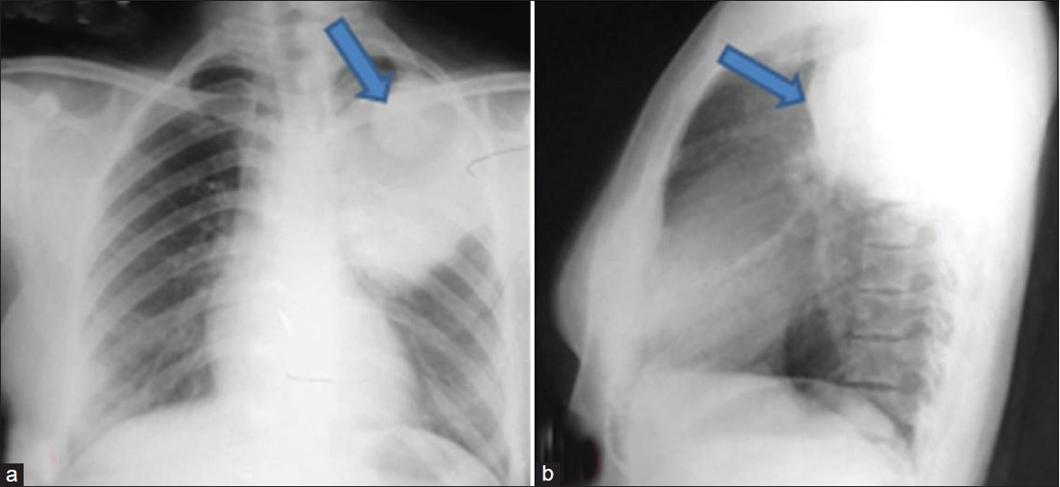
Chest X-ray (a) posterior anterior (PA) and (b) lateral views show well-defined homogeneous soft tissue opacity within posterior mediastinum, causing erosion of the 4th rib posteriorly, scalloping of the superior margin of the 5th rib and widening of the 4th intercostal space (arrow).
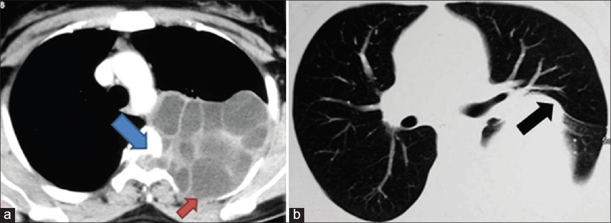
Figure 1
Chest X-ray (a) posterior anterior (PA) and (b) lateral views show well-defined homogeneous soft tissue opacity within posterior mediastinum, causing erosion of the 4th rib posteriorly, scalloping of the superior margin of the 5th rib and widening of the 4th intercostal space (arrow).
Contrast enhanced computed tomography (CT) thorax in (a) mediastinal and (b) lung windows show multiloculated cystic lesion in left paravertebral space, causing erosion of pedicle and lamina of the 4th dorsal vertebra, with intraspinal extension (blue arrow). The 4th rib is eroded (red arrow). Lung parenchyma is extrinsically compressed, unremarkable otherwise (black arrow).
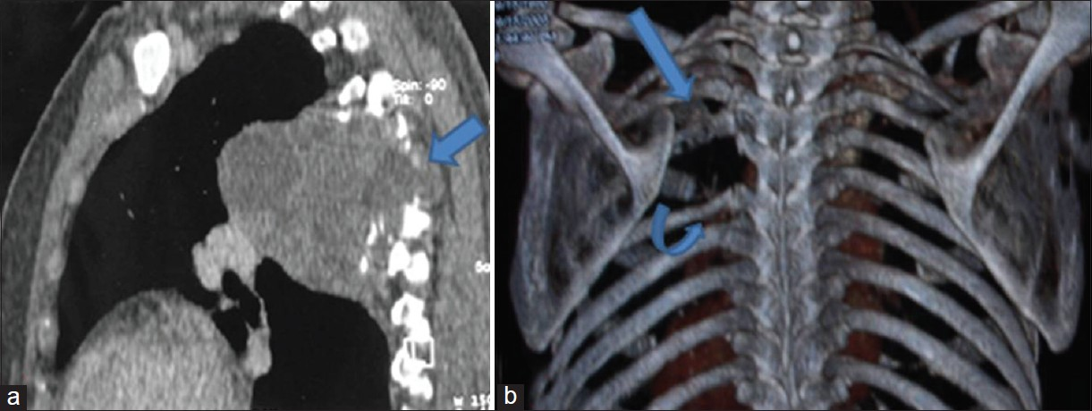
Figure 2
Contrast enhanced computed tomography (CT) thorax in (a) mediastinal and (b) lung windows show multiloculated cystic lesion in left paravertebral space, causing erosion of pedicle and lamina of the 4th dorsal vertebra, with intraspinal extension (blue arrow). The 4th rib is eroded (red arrow). Lung parenchyma is extrinsically compressed, unremarkable otherwise (black arrow).
(a) Three dimensional sagittal multiplanar reformation shows posterior mediastinal mass causing destruction of adjoining vertebra (arrow) with intraspinal extension. (b) Volume rendered coronal image shows erosion of the 4th rib (straight arrow) and scalloping of the 5th rib (curved arrow) with widened 4th intercostal space.
Figure 3
(a) Three dimensional sagittal multiplanar reformation shows posterior mediastinal mass causing destruction of adjoining vertebra (arrow) with intraspinal extension. (b) Volume rendered coronal image shows erosion of the 4th rib (straight arrow) and scalloping of the 5th rib (curved arrow) with widened 4th intercostal space.

Based on the imaging findings, the possible differential diagnosis included neurogenic tumors, aggressive malignant masses like round cell tumors (Ewing's sarcoma, primitive neuroectodermal tumors, and others), lymphoma, or metastasis. Since the mass was predominantly cystic with multiloculated appearance and considering the occupation of the patient, hydatid cyst was also considered a possible differential diagnosis.

The serological test with enzyme linked immunosorbant assay (ELISA) was positive for echinococcosis, thus suggesting hydatid cyst as a possible etiology. The patient was operated and the cystic mass along with the involved 4th rib were completely excised. The diagnosis was confirmed macroscopically preoperatively and by histopathological studies postoperatively. Postoperative recovery was uneventful and the patient was discharged and put on adjuvant chemotherapy with mebendazole for 2 years. The patient was monitored with routine follow-ups for 5 years with no evidence of recurrence.

DISCUSSION

Hydatid disease is a zoonosis caused by the parasite Echinococcus. There are two species of Echinococcus—Echinococcus granulosus and Echinococcus multilocularis. E. granulosus is most frequently encountered in hydatid disease in humans. Hydatid disease continues to be a major health problem in underdeveloped areas where animal husbandry is common and no veterinary control exists. It is endemic in parts of South America, Mediterranean regions, the Middle East, Africa, and Australia. Dogs or other carnivores are definitive hosts. Sheep and other ruminants are intermediate hosts. Humans are secondary hosts and are infected by the ingestion of food or water contaminated by eggs of these parasites.

In humans, the liver is the most frequent site of involvement followed by the lungs. Other sites in decreasing frequency include peritoneum, kidney, brain, mediastinum, heart, bone, soft tissues, spinal cord, spleen, pleura, adrenal glands, bladder, ovary, scrotum, and thyroid gland.

Mediastinal hydatid cysts are very rare, accounting for less than 0.1% of all cases and less than 1% of thoracic involvement. These occur more frequently in the posterior mediastinum.[1] The patients may remain asymptomatic, the lesion being discovered incidentally on a routine chest radiograph. The patients may present with non-specific symptoms of chest pain, cough, and dyspnoea. These symptoms may be produced as a result of involvement of neighboring structures and other etiologies like hemoptysis, superior vena cava syndrome, phrenic or recurrent laryngeal nerve compression, vertebral destruction with intraspinal extension and neurological symptoms, and Bernard–Horner syndrome.[2] The complications of mediastinal hydatid cyst can be significant and include rupture into the mediastinum, pleural cavity and right ventricle, cysto-aortic fistula with possible embolization, and infection and compression of vital structures.

Posterior mediastinal hydatid cyst with intraspinal extension is an extremely rare entity, with less than 10 similar cases reported in the literature till date.[34] Such masses with both intraspinal and extraspinal components assume a dumbbell shape.[5] Though the most common cause of dumbbell masses is neurogenic tumor, a large variety of unusual lesions may produce similar picture. These include neoplastic lesions such as benign/malignant peripheral nerve sheath tumors, plasmacytoma, chondrosarcoma, chondroid chordoma, superior sulcus tumor, metastasis, and nonneoplastic lesions such as infectious process (tuberculosis, hydatid cyst), aneurysmal bone cyst, synovial cyst, traumatic pseudomeningocele, arachnoid cyst, vertebral artery tortuosity, and hemangiomas. Paravertebral masses with vertebral destruction and intraspinal extension have been described as spinal hydatids by some authors,[6] and belong to Group 5, paravertebral hydatid disease, according to the Braithwaite and Lee's classification.[7]

Mediastinal echinococcosis is clinically and radiologically indistinguishable from other mediastinal cystic lesions. The differential diagnosis of posterior mediastinal cystic lesions includes bronchogenic cysts, cystic lymphangioma, enteric cyst, and neurogenic tumors. Presence of a posterior mediastinal mass with rib and vertebral destruction and intraspinal extension can raise the possibility of aggressive malignant masses like round cell tumor (Ewing's sarcoma, primitive neuroectodermal tumor, etc.), lymphoma, metastasis, or neurogenic tumor.[8]

Serological tests can often be negative, especially when the cyst is intact and uncomplicated. In suspected cases, aspiration cytology under imaging guidance is considered the procedure of choice for establishing the diagnosis.[9]

Radiological imaging consists of chest radiograph and CT of the thorax. CT defines the cystic nature of the mass and delineates its exact anatomical relations. Though presence of multiloculated appearance and calcification may suggest the diagnosis, there is no pathognomonic sign of hydatid cyst on CT. Magnetic resonance imaging is indicated in defining the intraspinal extension, especially when patients present with neurological signs.

The Gold Standard for treatment of mediastinal hydatid cyst is radical removal of the germinative membrane and pericyst.[10] If total excision is not possible due to invasion of vital structures, partial pericystectomy after removal of germinative membrane can be performed. It has been proposed that better results are obtained by combining surgical procedure with antihelminthic therapy postoperatively. World Health Organization recommends adjuvant chemotherapy with mebendazole or albendazole for 2 years postoperatively.

CONCLUSION

To conclude, hydatid cyst should always be considered in the differential diagnosis of mediastinal cystic lesions, especially in endemic areas, even when the lesion appears aggressive clinically and on imaging.

Source of Support: Nil

Conflict of Interest: None declared.

REFERENCES

  1. , , , , . Hydatid cyst: An unusual disease of the mediastinum. Acta Chir Belg. 2001;101:283-6.
    [Google Scholar]
  2. , , , , , . Primary hydatid cyst of the posterior mediastinum. Asian Cardiovasc Thorac Ann. 2007;15:e60-2.
    [Google Scholar]
  3. , , , , , . Posterior mediastinal paravertebral hydatid cyst presenting as spinal compression. A case report. Clin Neurol Neurosurg. 1990;92:149-51.
    [Google Scholar]
  4. , , , , , , . Rare location of a hydatid cyst- in the upper mediastinum migrating into the spinal channel. Rev Med Chir Soc Med Nat Iasi. 2001;105:573-5.
    [Google Scholar]
  5. , , , , , , . Unusual dumbbell tumours of the mediastinum and thoracic spine. J Clin Neurosci. 2006;13:958-62.
    [Google Scholar]
  6. , , . Dumbbell hydatid cyst of the spine: Case report and review of the literature. Spine (Phila Pa 1976). 2000;25:1296-9.
    [Google Scholar]
  7. , , . Vertebral hydatid disease: Radiological assessment. Radiology. 1981;140:763-6.
    [Google Scholar]
  8. , . Extrapleural extraosseous costal echinococcosis. Br J Radiol. 1979;52:918-9.
    [Google Scholar]
  9. , , , . Ultrasound guided fine-needle aspiration cytology: Diagnosis of hydatid disease of the abdomen and thorax. Diagn Cytopathol. 1995;12:173-6.
    [Google Scholar]
  10. , , , , , , . Mediastinal hydatid cysts: 8 cases. Arch Bronconeumol. 2000;36:221-4.
    [Google Scholar]

Fulltext Views
8,428

PDF downloads
3,063
View/Download PDF
Download Citations
BibTeX
RIS
Show Sections