Translate this page into:
Idiopathic Thrombus in the Common Carotid Artery on Digital Subtraction Angiography
Address for correspondence: Dr. Anand Alurkar, Department of Neurointervention, King Edward Memorial Hospital, Pune, India. E-mail: anandalurkar@gmail.com
-
Received: ,
Accepted: ,
This is an open-access article distributed under the terms of the Creative Commons Attribution License, which permits unrestricted use, distribution, and reproduction in any medium, provided the original author and source are credited.
This article was originally published by Medknow Publications & Media Pvt Ltd and was migrated to Scientific Scholar after the change of Publisher.
Abstract
In the present study, we discuss the accuracy of digital subtraction angiography (DSA) in diagnosis of thrombus in the common carotid artery and its role in the medical management of this disorder. Between 2006 and 2011, four patients (age group ranging from 26 to 48 years) presented to our institution with symptoms of stroke. DSA in all these patients showed cigar-shaped filling defect in the common carotid artery. All the patients were managed successfully with anticoagulation treatment. Follow-up Duplex scan was done in all the patients. DSA is the gold standard to diagnose free floating thrombus in the common carotid artery. Medical management can be effective in these patients but a multidisciplinary team approach is needed for appropriate management.
Keywords
CCA
idiopathic thrombus
stroke
INTRODUCTION

Mural thrombus in the common carotid artery is a rare entity and the etiology remains unclear. In this article, we discuss the importance of DSA in detecting thrombus in the CCA in four patients. On DSA, a cigar shaped filling defect in the common carotid artery suggestive of thrombus was seen in all four cases presented in this series. Detailed investigative evaluation was done to find any etiological and clinical association. We did not find any underlying etiology for the thrombus in these cases. All the patients were treated with anticoagulation medication. Literature review of CCA thrombus presenting with stroke suggested that this is an uncommon presentation.
CASE REPORT
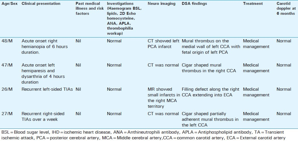
Four male patients (age group 26 to 48 years; mean age of 37 years), presented to our Institution with an abrupt onset of stroke. Detailed medical history, clinical examination, and investigative evaluation was done to evaluate the etiology of the thrombus in these relatively young patients. As no causative agent was found we concluded that these were ‘idiopathic’ cases. Digital subtraction angiogram was performed in the patients. It revealed mural thrombus [Figures 1–4] in the common carotid artery in all the cases. Findings are summarized in [Table 1]. We did not find any etiological association for the thrombus. A complete clinical evaluation including a hemogram, 2D Echocardiogram, and measurement of blood sugar level, lipids, serum homocysteine, antineutrophilic antibody, and anti-phospholipid antibody levels were done to rule out a hypercoagulable state. All the measured parameters were within normal limits. The patients were treated with Enoxaparin injection (0.6 mg subcutaneous twice a day) followed by Warfarin to maintain international normalized ratio of 2-2.5. All four patients improved with anticoagulation medication and were discharged with no neurological deficit on the 7th day. Surgical intervention was not required in any of our cases since medical management was effective in all the cases. Duplex Doppler scan done after 6 months in all the patients [Figure 5] showed normal flow and complete clot dissolution without any underlying wall abnormalities like atheromatous plaque or dissection.
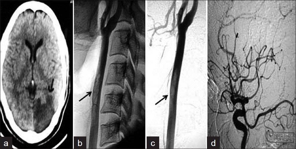
- Patient 1 (a) CT scan shows infarct in the left PCA territory (arrow) and (b, c) angiogram shows the cigar-shaped thrombus (arrows) in the left CCA. (d) Angiogram (lateral view) shows the fetal PCA.
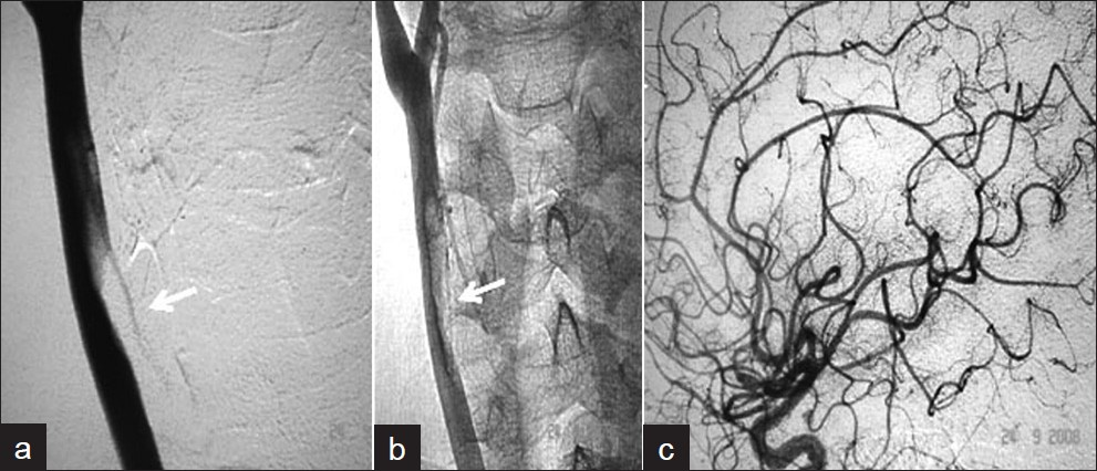
- Patient 2 (a, b) Right CCA angiogram shows the filling defect suggestive of mural thrombus and (c) normal intracranial cerebral circulation.
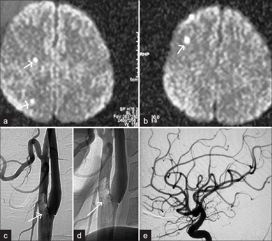
- Patient 3 (a, b) MR shows small foci of restricted diffusion suggesting acute infarcts in the right MCA territory (arrows). Patient 3 (c,d) Right CCA angiogram on lateral views shows filling defect (arrows) in the distal common carotid artery extending into the external carotid artery. (e) Intracranial cerebral angiogram is normal.
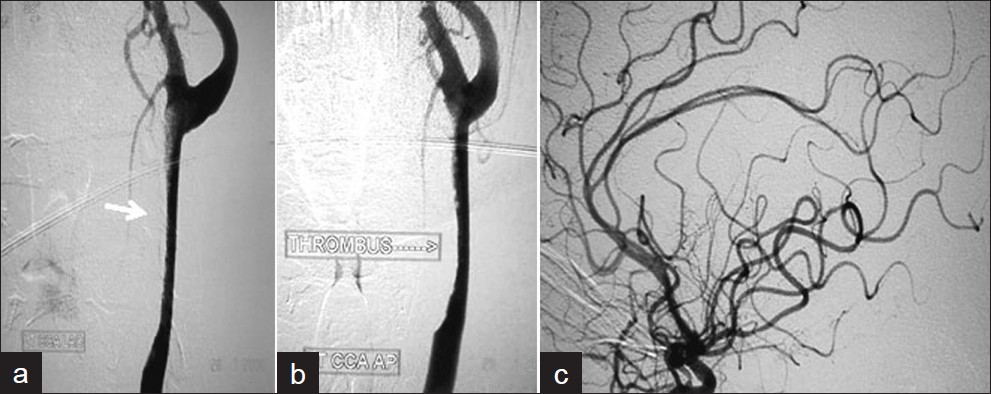
- Patient 4 (a,b) Left CCA angiogram shows cigar-shaped partially adherent mural thrombus in the left CCA and (c) normal intracranial circulation.
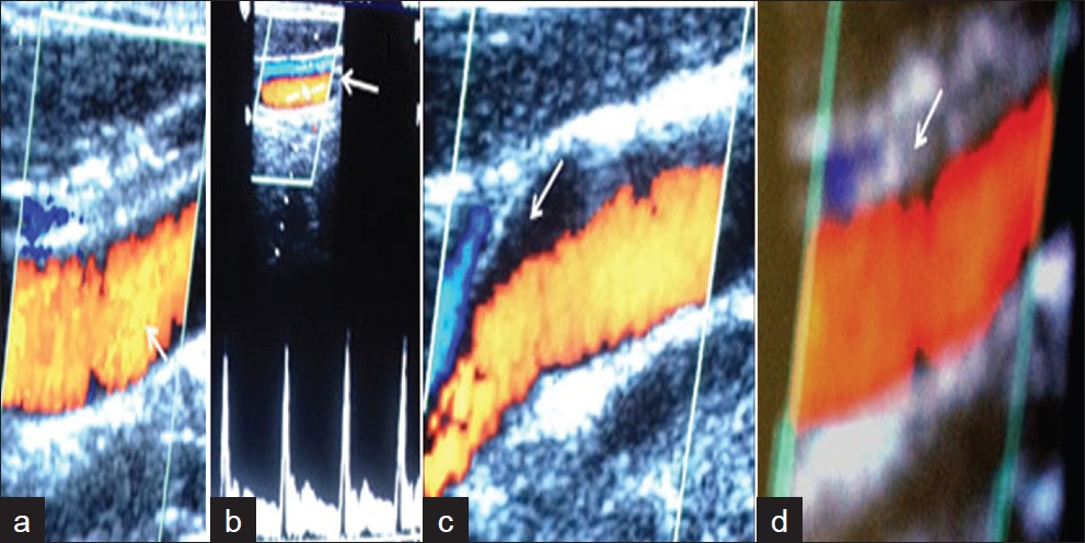
- Follow-up carotid Doppler images of (a) Patient 1 (b) Patient 2 (c) Patient 3 and (d) Patient 4 show complete dissolution of thrombus with normal flow in the artery.
DISCUSSION
The first case of CCA thrombus diagnosed by conventional angiography was reported by Akins et al.,[1] in a patient with severe iron deficiency anemia and reactive thrombocytosis secondary to menorrhagia. Devuyst et al.,[2] described the ultrasonographic feature of this uncommon finding in young patients with stroke. The main concern in these patients was risk of recurrent embolic events. On survey of literature about the idiopathic thrombus occurrence in the CCA we found a few reports.[34] Bhatti et al.,[5] in their review found a total of 145 cases of free floating thrombus in the carotid axis of which 75% were in the ICA with a male predominance and more common in younger age groups. Controversial options for treatment of carotid thrombus have been discussed in detail in the literature by Hill et al.[6] Ferraro et al.[7] in their series of 16 patients discussed about the role of various imaging modalities for the diagnosis of free floating thrombus (FFT) and analyzed the efficacy of carotid endarterectomy in these patients. These authors also described a rare case of FFT on residual carotid intimal flap after carotid endarterectomy in a 75-year-old man, who was subsequently treated with thromboendarterectomy.[8] They also reported the first case of emergency thrombectomy in ICA occlusion in a patient with malignant mesothelioma.[9] In all the four patients of our present case series, immediate surgery was deferred as per the decision of the surgical team, due to the location of the thrombus (exclusively within the CCA) and high risk of stroke associated with the surgery. Based on the recommendations of our multidisciplinary team, a decision was taken to medical manage the patients with anticoagulation medication.
CONCLUSION
DSA is the gold standard to diagnose the mural thrombus in the common carotid artery. In the present series the etiology of thrombus was unclear after extensive detailed clinical and imaging evaluation. Medical management can be effective in selected cases and a multimodality approach is needed in deciding the therapeutic strategy.
Source of Support: Nil
Conflict of Interest: None declared.
Available FREE in open access from: http://www.clinicalimagingscience.org/text.asp?2012/2/1/20/95434
REFERENCES
- Carotid artery thrombus associated with severe iron-deficiency anemia and thrombocytosis. Stroke. 1996;27:1002-5.
- [Google Scholar]
- Focal adherent thrombus in the common carotid artery: Clinical, ultrasonographic, and pathogenic aspects in two cases. J Ultrasound Med. 2000;19:707-11.
- [Google Scholar]
- Idiopathic floating thrombus of the common carotid artery: Diagnosis and treatment options. J Ultrasound. 2010;13:57-60.
- [Google Scholar]
- Idiopathic free-floating thrombus of the common carotid artery. Can J Neurol Sci. 2002;29:97-9.
- [Google Scholar]
- Free-floating thrombus of the carotid artery: Literature review and case reports. J Vasc Surg. 2007;45:199-205.
- [Google Scholar]
- Extensive mobile thrombus of the internal carotid artery: A case report, treatment options, and review of the literature. Am Surg. 2005;71:853-5.
- [Google Scholar]
- Free-floating thrombus in the internal carotid artery: Diagnosis and treatment of 16 cases in a single center. Ann Vasc Surg. 2011;25:805-12.
- [Google Scholar]
- Free floating thrombus developed on a residual carotid intimal flap after carotid endarterectomy. Diagnosis and treatment. 2010;24:573-4.
- [Google Scholar]
- Mesothelioma and internal carotid artery occlusion: Acute ischemic stroke and efficacy of emergency carotid thrombectomy. Ann Vasc Surg. 2010;24:257.
- [Google Scholar]


