Translate this page into:
Calcified Triticeous Cartilage Detected on Digital Panoramic Radiographs in a Sample of Lebanese Population
Address for correspondence: Dr. Georges Aoun, Department of Oral Medicine and Maxillofacial Radiology, Faculty of Dental Medicine, Lebanese University, Beirut, Lebanon. E-mail: aoungeorges@yahoo.com
-
Received: ,
Accepted: ,
This is an open access journal, and articles are distributed under the terms of the Creative Commons Attribution-NonCommercial-ShareAlike 4.0 License, which allows others to remix, tweak, and build upon the work non-commercially, as long as appropriate credit is given and the new creations are licensed under the identical terms.
This article was originally published by Medknow Publications & Media Pvt Ltd and was migrated to Scientific Scholar after the change of Publisher.
Abstract
Objective:
Triticeous cartilage is a small ovoid structure belonging to the laryngeal skeleton. When calcified, it becomes visible on panoramic radiographs and be mistaken for a carotid artery calcification (CAC) associated with cerebrovascular accidents. This study aimed to estimate the prevalence of calcified triticeous cartilage (CTC) detected by means of digital panoramic radiographs in a sample of Lebanese population.
Materials and Methods:
Digital panoramic radiographs of 500 Lebanese adult patients (281 females and 219 males) with a mean age of 47.9 years were included in this study and examined for CTC. The IBM® SPSS® for Windows version 20.0 (SPSS, Chicago, IL, USA) was used to carry out statistical analysis of the data collected.
Results:
Nearly 10.6% (53 out of 500) of the radiographs examined presented CTC. Of all the calcifications, 11 were on the right side, 5 on the left side, and 37 were bilateral. The cases detected belonged to 31 females and 22 males with an average age of 55.6 years (ranging from 24 to 85 years). Chi-square test did not show any statistical connection between gender and CTC, while Spearman's correlation analysis showed low positive correlation with age (r = 0.146).
Conclusion:
CTC can be detected on panoramic radiographs taken in daily dental practice; its identification is essential to avoid misdiagnosis with other calcifications in the neck region closely related to life-threatening risks such as CAC.
Keywords
Calcification
Lebanese
panoramic radiography
population
triticeous cartilage

INTRODUCTION
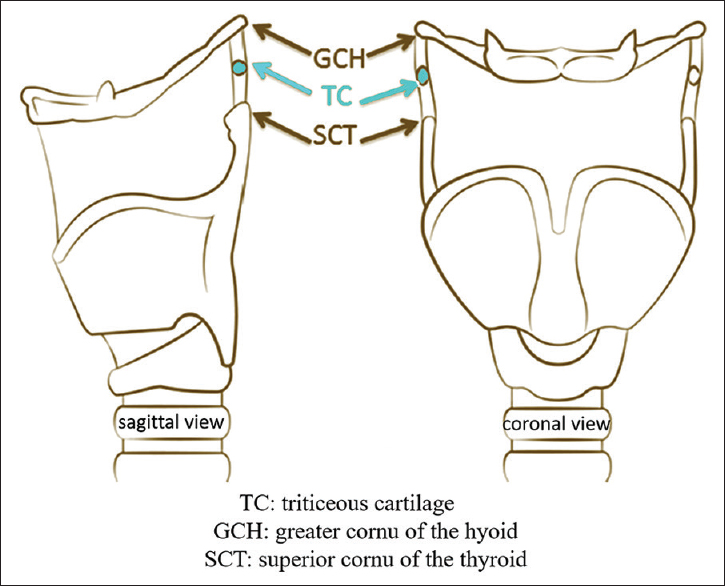
Triticeous cartilage is a small ovoid cartilaginous structure located within the thyrohyoid ligament extending from the hyoid bone (distal aspect of the greater cornu) to the thyroid cartilage (tip of the superior cornu) at the level of C3 and C4 vertebrae[123] [Figure 1].

- Schema illustrating the thyrohyoid ligament and the triticeous cartilage. (TC: Triticeous cartilage; GCH: Greater cornu of the hyoid bone; SGT: Superior cornu of the thyroid cartilage).
The role of the triticeous cartilage is unidentified; according to many researchers, it may serve to reinforce the thyrohyoid ligament.[2345]
In some cases, just like other structures of the laryngeal skeleton, the triticeous cartilage undergoes calcification and becomes visible on panoramic radiographs, especially when the visual field extends inferiorly.[3]
In dentistry, the calcified triticeous cartilage (CTC) represents for the practitioners a major challenge when detected radiologically. Due to its location, it may be mistaken for a carotid artery calcification (CAC) closely related to cerebrovascular accidents.[23] However, although sharing the same radiological location, these two entities present different shapes making their diagnosis less problematic; in fact, CTC is generally oval, surrounded by a smooth, well-defined border, while CAC is mostly verticolinear with irregular margins and areas of radiolucencies.[2367]
With the absence of any radiologic investigation for CTC in Lebanon, the purpose of this study was to evaluate these types of calcifications in a Lebanese sample by means of digital panoramic radiographs.
MATERIALS AND METHODS
This was a retrospective study conducted on 500 archived digital panoramic radiographs of Lebanese adults (219 males and 281 females) taken originally for oral and dental diagnostic reasons in a specialized maxillofacial imaging center in Beirut, Lebanon.
As by the center rules, all patients gave their consent systematically for anonymous future use of their radiographs for research aims.
All images were acquired by the Pax Zenith digital panoramic unit (Vatech, Korea). The settings of the unit were set to 60–90 kV and 6–10 mA; exposure time was 10–20 s.
Exclusion criteria comprised the absence of any information about patients' age and gender and radiographs with bad quality and/or not showing C3–C4 vertebrae.
The investigator was an experienced (more than 15 years) oral and maxillofacial radiologist; he assessed the images on the same monitor over four sessions spaced by a 20-day period. To reduce the errors, 100 images were randomly chosen and rechecked 15 days later.
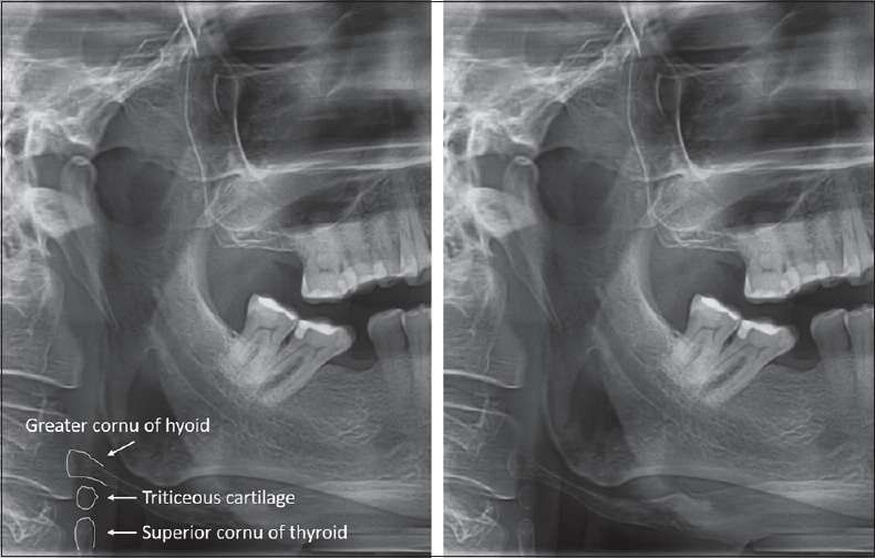
The CTC was identified as an oval with a smooth, well-defined radiopacity located between the superior horn of the thyroid cartilage and the greater horn of the hyoid bone at the level of the third and the fourth cervical vertebrae [Figure 2].

- A radiograph in the region of C3–C4 vertebrae showing the calcified triticeous cartilage as considered by our study.
Data were analyzed by means of IBM® SPSS® for Windows, version 20.0 (SPSS, Chicago, IL, USA). Descriptive statistics of age, gender, and CTC were elaborated. Chi-square test and Spearman's correlation analysis were performed. Statistical significance was set at 0.05.
RESULTS
Of the 500 digital panoramic radiographs, 281 and 219 were, respectively, of females and males. The patients' age ranged between 18 and 88 years with a mean of 47.9 years.
In our study population, CTC was seen in 53 cases (10.6%) (31/281 women and 22/219 men); among these, 11 (2.2%) (4 women and 7 men) were on the right side, 5 (1%) (3 women and 2 men) on the left, and 37 (7.4%) (24 women and 13 men) were bilateral [Graph 1].

- Frequency of calcified triticeous cartilage (0 denotes absence, 1 denotes right side, 2 denotes left side, and 3 denotes bilateral formation).
When assessing the association between gender and CTC by Chi-square test, no statistically significant difference was found (P = 0.7).
In our sample, the mean age of patients presenting CTC was 55.6 years (ranging from 24 to 85 years) [Graph 2].

- Association of calcified triticeous cartilage with age.
Spearman's correlation analysis showed a low positive correlation between age and CTC (r = 0.146).
DISCUSSION
Soft-tissue calcifications within the head-and-neck regions are commonly found in patients seeking dental treatments. Generally asymptomatic, they may be accidentally detected on panoramic radiographs. Among these calcifications is CTC, which is usually unknown by dentists and could be radiologically mistaken for CAC, a dangerous condition potentially leading to cerebrovascular accidents.[89]
In the literature, a small number of studies investigated CTC in different ways and populations. Ajmani et al.[1011] assessed it as a part of the thyroid ligament on Indian and Nigerian adults' cadavers, while Alqahtani et al.[1] and Hatleyet al.[12] used, respectively, computed tomography technique and neck radiographs to describe its appearance.
In our study conducted on Lebanese adults using digital panoramic radiographs, the incidence of CTC which was found to be 10.6% cannot be compared with any of the findings from the above-cited studies due to the difference in the assessment methods. However, this result was slightly higher than what was reported by Ahmad et al.[2] (8.6%) who used the same technique. This small difference may be due to the dissimilarity in the sample sizes (n = 500 vs. n = 847).
In our sample, we found a low positive correlation between age and CTC, with an average of 55.6 years. Although our result agrees with what was stated by Ahmad et al.[2] (51–60 years), a comparison cannot be made as the age group investigated by the authors (40 years and older) is different from ours (ranging from 18 to 88 years).
On the other hand, contrary to Ahmad et al.'s[2] report of female predominance, there was no significant connection between CTC and gender in our study (P = 0.7).
Finally, our study aiming to estimate the prevalence of CTC in a sample of Lebanese population is not without limitation. Although radiologic evaluations were made with a high reliability, the restricted number of radiographs assessed makes necessary the investigation on a larger group, which can lead to more precise results.
CONCLUSION
CTC may be accidentally noticed on panoramic radiographs taken in dental practice. Clinicians should think about this entity when a well-defined oval radiopacity is seen at the level of the third and the fourth cervical vertebrae.
Financial support and sponsorship
Nil.
Conflicts of interest
There are no conflicts of interest.
Available FREE in open access from: http://www.clinicalimagingscience.org/text.asp?2018/8/1/16/230278
REFERENCES
- Triticeous cartilage CT imaging characteristics, prevalence, extent, and distribution of ossification. Otolaryngol Head Neck Surg. 2016;154:131-7.
- [Google Scholar]
- Triticeous cartilage: Prevalence on panoramic radiographs and diagnostic criteria. Oral Surg Oral Med Oral Pathol Oral Radiol Endod. 2005;99:225-30.
- [Google Scholar]
- Discrimination between calcified triticeous cartilage and calcified carotid atheroma on panoramic radiography. Oral Surg Oral Med Oral Pathol Oral Radiol Endod. 2000;90:108-10.
- [Google Scholar]
- Anomalies and alterations of the hyoid-larynx complex in forensic radiographic studies. Am J Forensic Med Pathol. 2004;25:14-9.
- [Google Scholar]
- Considerations in detecting soft tissue calcifications on panoramic radiography. J Int Oral Health. 2016;8:742-6.
- [Google Scholar]
- Soft tissue calcified in mandibular angle area observed by means of panoramic radiography. Int J Clin Exp Med. 2014;7:51-6.
- [Google Scholar]
- Carotid calcification on panoramic radiographs: An important marker for vascular risk. Oral Surg Oral Med Oral Pathol Oral Radiol Endod. 2002;94:510-4.
- [Google Scholar]
- Carotid artery calcification: A digital panoramic-based study. Diseases. 2018;6 pii: E15
- [Google Scholar]
- A metrical study of laryngeal cartilages and their ossification. Anat Anz. 1980;148:42-8.
- [Google Scholar]
- A metrical study of the laryngeal skeleton in adult Nigerians. J Anat. 1990;171:187-91.
- [Google Scholar]
- The pattern of ossification in the laryngeal cartilages: A radiological study. Br J Radiol. 1965;38:585-91.
- [Google Scholar]


