Translate this page into:
Masseter Muscle Thickness in Unilateral Partial Edentulism: An Ultrasonographic Study
Address for correspondence: Dr. S. Sathasivasubramanian, No. 1, Sri Ramachandra Nagar, Porur, Chennai - 601 116, Tamil Nadu, India. E-mail: dr_sathasivam@yahoo.co.in
-
Received: ,
Accepted: ,
This is an open access article distributed under the terms of the Creative Commons Attribution-NonCommercial-ShareAlike 3.0 License, which allows others to remix, tweak, and build upon the work non-commercially, as long as the author is credited and the new creations are licensed under the identical terms.
This article was originally published by Medknow Publications & Media Pvt Ltd and was migrated to Scientific Scholar after the change of Publisher.
Abstract
Introduction:
Teeth and facial muscles play a very important role in occlusal equilibrium and function. Occlusal derangement, seen in unilateral partially edentulous individuals, has an effect on masseter muscle anatomy and function. The present study aims to evaluate masseter muscle thickness in unilateral partial edentulism.
Patients and Methods:
Institutional ethics committee approval was obtained before the commencement of the study. The study involved patients who routinely visited the Department of Oral Medicine and Radiology, Sri Ramachandra University. The study sample included 27 unilateral edentulous patients (Group E) and 30 controls (Group C). The masseter muscle thickness was evaluated using high-resolution ultrasound real-time scanner (linear transducer − 7.5–10 MHz) at both relaxed and contracted states.
Statistical Analysis Used:
The results were analyzed using paired t-test and independent t-test. Duration of edentulism and muscle thickness was assessed using Pearson's correlation coefficient.
Results:
The study patients’ age ranged between 25 and 48 years (mean – 36 years). The comparative evaluation of masseter muscle thickness between the dentulous and edentulous sides of experimental group was statistically significant (P < 0.05). However, no statistically significant difference in masseter muscle thickness was found between the dentulous side of control and experimental groups. The correlation between the duration of partial edentulism and muscle thickness was statistically insignificant.
Conclusion:
The study proves masseter atrophy in the edentulous side. However, since the difference is found to be marginal with the present sample, a greater sample is necessary to establish and prove the present findings as well as to correlate with the duration of edentulism. Further studies are aimed to assess the muscle morphology after prosthetic rehabilitation.
Keywords
Masseter
masseter muscle thickness
ultrasound
unilateral partial edentulism

INTRODUCTION
Muscles of mastication and teeth play an important role in maintaining the occlusal equilibrium.[1] Among the various muscles of mastication, masseter plays an important role in chewing action and hence is directly influenced by changes in the harmonious occlusion.[2] Muscle thickness has been considered one of the indicators of jaw muscle activity.[3] Various imaging techniques such as the ultrasound, computed tomography (CT), and magnetic resonance imaging (MRI) have been employed to evaluate and measure the muscle thickness.[456] Over the years, ultrasonography has proved to be a reproducible, simple, and inexpensive method for measuring muscle thickness accurately.[3]
Although various studies have been done regarding the relationship of masseter muscle and craniofacial morphology using ultrasonography,[7] the study on masseter muscle thickness in unilateral partially edentulous patients is very sparse. Till date, there is only one study in the literature to have evaluated the effect of unilateral partial edentulism on the thickness of masseter and anterior temporalis muscles.[1] Hence, the current study aims to evaluate the effect of unilateral partial edentulism on masseter muscle thickness using ultrasonography.
SUBJECTS AND METHODS
The study involved patients who routinely visited the Department of Oral Medicine and Radiology, Faculty of Dental Sciences, Sri Ramachandra University, for various teeth-related and oral mucosal diseases. Institutional ethics committee approval (CSP/14/APR/34/39) was obtained before the commencement of the study. The study sample included 27 unilateral edentulous patients (Group E) and 30 controls (Group C). The experimental group (Group E) included patients with either partially edentulous maxilla (missing 15, 16, 17/25, 26, and 27) or partially edentulous mandible (missing 45, 46, 47/35, 36, and 37). Patients with bilateral partial edentulism were excluded from the study. The control group included age- and gender-matched individuals with no evidence of missing teeth. The masseter muscle thickness was evaluated using high-resolution ultrasound real-time scanner at both relaxed and contracted states at the Department of Radiology and Imaging sciences, Sri Ramachandra Medical Centre, Chennai. To eliminate the interobserver difference, the same operator evaluated the masseter thickness in each study individual, using a real-time scanner with a linear transducer in the range of 7.5–10 MHz (GE LOGIQP5, GE Health Care, UK). The operator applied a water-based gel to the probe and adequate pressure was applied to the probe before the imaging procedure as shown in Figure 1. To produce the strongest echo from the mandibular ramus, the angle of probe was adjusted such that the scan plane was perpendicular to its surface. The patients were instructed to bring their teeth at centric occlusion and lightly touch their teeth on the dentulous side for experimental and control groups. They were then demonstrated and instructed to do maximal clenching, which was done repeatedly for few times. The ultrasound findings were recorded at moderate clenching which was intermediate between slight tooth contact and maximal clenching. Contrast between muscle and subcutaneous tissue was enhanced by asking the participant to clench and relax alternately. The thickest part of the masseter which was close to the level of the occlusal plane, approximately in the middle of the mediolateral distance of the ramus, was measured. The study participants were placed in the supine position and bilateral measurements were taken when the teeth were made to occlude gently with the muscles in a relaxed position and when the muscles contracted during maximal clenching. During the time of scanning and image acquisition, the measurements were recorded in centimeters (cm).

- Ultrasonic probe used in the measurement for the masseter muscle.
Statistical methods
Data of muscle thickness in cm were recorded and statistical analyses were performed using SPSS software version 18 (SPSS Inc. Released 2009. PASW Statistics for Windows, Version 18.0. Chicago: SPSS Inc.). The muscle thickness between edentulous and dentulous sides of Group E was compared using paired t-test. The muscle thickness in the dentate side of Group E and controls (Group C) was compared using independent t-test. Duration of edentulism and muscle thickness was assessed using Pearson's correlation coefficient.
RESULTS
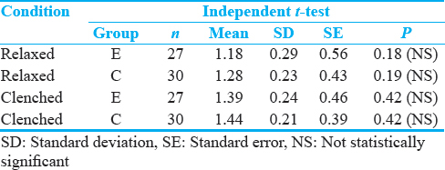
In the experimental group (n = 27), the participants’ age ranged between 25 and 48 years, with a mean age of 36 years. In the control group (n = 30), the age range was found to be in the range of 26–50 years, with a mean age of 37 years. Among the experimental group, the number of males was found to be 13 and females to be 14, and in the control group, there was an equal number of males and females (15). The duration of edentulism ranged between 6 months and 10 years, with a mean value of 5 years. The masseter muscle thickness in relaxed and contracted states in both experimental and control groups is illustrated in Table 1. In Group E, the masseter muscle thickness in relaxed position ranged from 0.74 to 1.69 cm (mean: 1.08 cm) and in contracted position ranged from 0.92 to 1.96 cm (mean: 1.23 cm) in the edentulous side, and the thickness of the muscle in relaxed position ranged from 0.82 to 1.88 cm (mean: 1.23 cm) and in contracted position ranged from 0.92 to 1.99 cm (mean: 1.39 cm) in the dentulous side. In Group C, the thickness of the muscle in relaxed position ranged from 0.87 to 1.75 cm (mean: 1.27 cm) and in contracted position ranged from 0.97 to 1.94 cm (mean: 1.44 cm) [Figures 2 and 3].


- Ultrasonographic image of left and right masseter muscle thickness in both relaxed and contracted positions in age- and gender-matched controls (Group C).

- Ultrasonographic image of left and right masseter muscle thickness in both relaxed and contracted positions in unilateral partially edentulous patients (Group E).
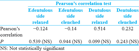
The difference in the masseter muscle thickness between the dentulous and partially edentulous sides in experimental group in both relaxed and contracted positions was statistically significant (P < 0.05) [Table 2]. However, the difference of masseter muscle thickness between the dentulous side of experimental and the control groups was not statistically significant (P > 0.05) [Table 3]. The correlation between duration of edentulism and muscle thickness was found to be statistically insignificant (P > 0.05) [Table 4].


DISCUSSION
Teeth and muscles work in harmony to produce an occlusal equilibrium.[1] However, such equilibrium is lost when teeth or muscle of mastication deviate from the normal physiology and function. During conditions when teeth are lost, people tend to chew on side which has the greatest number of teeth contacts during lateral gliding movements and because of which the masticatory load is not equally distributed bilaterally.[8] Hence, the bilateral and unilateral edentulous people experience difficulty in maintaining an effective chewing function. The unilateral partially edentulous patients prefer to use the dentate side more frequently because of the greater number of tooth contacts.[1] As the duration of edentulousness increases, and with no effective chewing occurring on the edentulous side, there could be an adverse effect on the masseter which is supposed to play a major role in chewing function.[2] The discrepancies between the teeth and muscle function result in the loss of occlusal equilibrium.
The masseter muscle contributes to higher activity in the clenching effort and measurement of its thickness has been considered an indicator of jaw muscle function.[2] Hence, the evaluation and comparison of masseter muscle thickness in unilateral edentulous and dentulous patients indicate the changes occurring when there is loss of occlusal equilibrium. Although various investigative procedures such as CT, MRI, and ultrasonography have been used to evaluate the muscle thickness, ultrasonography was chosen since it was proven to be a rapid, easy procedure, which is a simple, reproducible, and comparatively less expensive method for soft-tissue imaging. It has been shown that ultrasound can be used to accurately measure muscle thickness, when a strict mentioned imaging protocol is observed by the operator.[3]
In this study, the examination was done in supine position for the participants in relaxed and clenching positions to measure the muscle thickness which is in accordance with the study done by Morse and Brown for masseteric hypertrophy.[9] In a study done by Roarket al.,[10] moderate level of clenching was achieved in the study participants which was half way or 50% effort of slight teeth contact and maximal clenching, the same of which was employed in the present study following which ultrasound findings were recorded. Muscle thickness was recorded in the relaxed position with minimal pressure given to the probe during examination. The thickness of the masseter muscle in contracted position was greater than in relaxed position which is attributable to the muscle physiology that in relaxed states the muscle tone is lower than that in the contracted state.[1]
In the present study, the thickness of masseter muscle in the unilateral partially edentulous side is lesser than in the dentulous side in the experimental group in both relaxed and contracted states, the difference of which was statistically significant. This could be probably correlated with the disuse atrophy of the muscle which occurred as a result of unilateral chewing on the dentate side after tooth loss. This result of the present study is in correlation with the results of study by Koca-Ceylan et al.,[1] who have also reported a similar difference but proved to be statistically insignificant. The authors have concluded that unilateral chewing habits and duration of partial edentulism do not affect the muscle thickness.[1] In a study done by Bhoyar et al., comparing masseter muscle thickness in completely edentulous patients, the thickness was significantly lower than that of the dentulous patients, which was attributed to the atrophy in muscles of mastication with loss of teeth.[11] The present study shows a significant difference in the masseter thickness within the same patient between dentulous and edentulous sides. The significant difference could be due to excessive use of dentate side for chewing and disuse atrophy on the edentulous side. Discrepancies between the muscle measurement values in this study and those found by other investigators may be due to disparities between the sample population, difference in the location point of the transducers, imaging modalities employed,[1] and the orientation and size of the muscle fibers that may have genetic and environmental backdrop.
Despite a suggested excessive chewing by the edentulous patients on the dentulous side, the difference in the masseter thickness of dentate side of edentulous patients and the dentate patients was insignificant. This may imply that when masseter exhibits a proper chewing efficiency, the muscle fibers tend to maintain their anatomy, function, and normal equilibrium. The correlation between the duration of edentulism and muscle thickness was found to be statistically insignificant, which necessitates a greater sample size to prove this observation. Moreover, studies to evaluate the masseter thickness correlation with the period of edentulism, in bilaterally partially edentulous and completely edentulous patients, will help in assessing the pattern of disuse atrophy of masseter among the edentulous patients.
CONCLUSIONS
The present study indicates disuse atrophy of the masseter muscle in the edentulous side. High-resolution ultrasound is found to be very useful in identifying the changes in masseter muscle thickness. However, a larger sample size may be required to correlate the present findings with the duration of edentulism and further studies are aimed to assess the masseter muscle thickness after prosthetic rehabilitation.
Declaration of patient consent
The authors certify that they have obtained all appropriate patient consent forms. In the form the patient(s) has/have given his/her/their consent for his/her/their images and other clinical information to be reported in the journal. The patients understand that their names and initials will not be published and due efforts will be made to conceal their identity, but anonymity cannot be guaranteed.
Financial support and sponsorship
Nil.
Conflicts of interest
There are no conflicts of interest.
Available FREE in open access from: http://www.clinicalimagingscience.org/text.asp?2017/7/1/44/221870
REFERENCES
- The effect of unilateral partial edentulism to muscle thickness. Saudi Med J. 2003;24:1352-9.
- [Google Scholar]
- Relationship between masseter muscle form and occlusal supports of remaining teeth. Kurume Med J. 2012;59:5-15.
- [Google Scholar]
- Masseter muscle thickness measured by ultrasonography and its relation to facial morphology. J Dent Res. 1991;70:1262-5.
- [Google Scholar]
- Relationship between the maxillofacial morphology and the masseter muscle. Orthod Waves. 2004;63:1-10.
- [Google Scholar]
- A morphological study of the masseter muscle using magnetic resonance imaging in patients with jaw deformity, Cases demonstrating mandibular deviation. J Jpn Stmatol Soc. 2006;55:17-22.
- [Google Scholar]
- Relationship between jaws and masseter muscle by superimposing MR images on the cephalogram. Stomatol Soc J. 2006;73:116-24.
- [Google Scholar]
- Relationship between masseter muscle size and maxillary morphology. Eur J Orthod. 2011;33:654-9.
- [Google Scholar]
- Ultrasonic diagnosis of masseteric hypertrophy. Dentomaxillofac Radiol. 1990;19:18-20.
- [Google Scholar]
- Effects of interocclusal appliances on EMG activity during parafunctional tooth contact. J Oral Rehabil. 2003;30:573-7.
- [Google Scholar]
- Effect of complete edentulism on masseter muscle thickness and changes after complete denture rehabilitation: An ultrasonographic study. J Investig Clin Dent. 2012;3:45-50.
- [Google Scholar]


