
Translate this page into:
Un-re-sheath-able Misaligned pCONus Device: Case Report of a Unique Complication
-
Received: ,
Accepted: ,
This is an open access article distributed under the terms of the Creative Commons Attribution-NonCommercial-ShareAlike 3.0 License, which allows others to remix, tweak, and build upon the work non-commercially, as long as the author is credited and the new creations are licensed under the identical terms.
This article was originally published by Medknow Publications & Media Pvt Ltd and was migrated to Scientific Scholar after the change of Publisher.
Abstract
pCONus is a stent like endovascular device which aids in retention of coils within wide necked bifurcation aneurysms. It is retrievable even after complete deployment and is detached electrolytically. pCONus aided coiling of wide necked bifurcation aneurysms has a high technical success rate and a good safety profile. Different complications have been described in literature with the usage of pCONus. This case report describes a yet unreported complication of un-re-sheath-ability of a misaligned deployed pCONus device.
Keywords
Aneurysm
endovascular coiling
pCONus device
waffle cone technique

Corrected and Republished
Corrected and Republished: Misaligned pCONus Device: Case Report of a Unique Complication
Correction to: J Clin Imaging Sci 2017, 7:29; DOI 10.4103/jcis.JCIS_36_17
Certain edits with which the article was published earlier was not acceptable to the first and corresponding author.
Hence, the article is being republished with approved changes from the first and corresponding author.

INTRODUCTION
Treatment of wide necked bifurcation aneurysms incorporating part of parent vessel has always been difficult.[12345] pCONus device is an improvised stent made for coiling wide necked bifurcation aneurysms using the “waffle cone” technique.[2] Studies on pCONus-assisted coiling have demonstrated a high technical success and a desirable safety profile.[14] Various technical and long-term complications have been observed.[145] Technical complication of a un-re-sheath-able misaligned deployed pCONus device has never been described.
CASE REPORT
A 64-year-old female came with complaints of sudden onset severe headache which had started 3 days earlier. There was no neurological deficit. Plain computed tomography scan of the brain showed Fisher Grade 1 subarachnoid hemorrhage in the right Sylvian fissure. Digital subtraction angiography through a right femoral access showed a wide necked middle cerebral artery bifurcation aneurysm measuring 7.3 mm × 5.8 mm, neck measuring 4 mm [Figure 1]. A decision to treat the aneurysm endovascularly with pCONus device assistance was made after an interdisciplinary discussion. Under adequate heparinization, the right femoral angiographic sheath was exchanged over a wire for a 7 French long sheath, and a 0.070” 6F Neuron guiding catheter (Penumbra Inc, Alameda, California, USA) was placed coaxially with its tip in the upper cervical segment of the internal carotid artery. A Vasco 0.021” microcatheter (Balt, Montmorency, France) was placed through the guiding catheter with its tip in the center of the aneurysm. A 20 mm long pCONus device with 6 mm crown was delivered through the microcatheter, the crown was deployed within the aneurysm and the microcatheter was pulled back to place the crown at the base of the aneurysm covering the neck. Four tablets of 75 mg prasugrel were administered through a nasogastric tube at this point. The rest of the device was deployed in the middle cerebral artery by unsheathing it. After unsheathing the device, there was prolapse of one of the crown petals into the inferior division of middle cerebral artery [Figure 2]. Repeated attempts at re-sheathing the device with the aim to reposition it by two operators failed. It was decided to proceed with coiling of the aneurysm with this misaligned position of the pCONus device rather than retrieve the device in its deployed state. A Vasco 0.010″ microcatheter (Balt, Montmorency, France) was used to intubate the aneurysm through the pCONus device, and the aneurysm was coiled progressively using multiple coils (a three-dimensional [3D] coil followed by soft coils) with frequent repositioning of prolapsing coil loops. After the aneurysm was near-totally filled and there was minimal residual filling, further coil loops prolapsed into the parent vessel relentlessly on repeated trying, and dislodged a loop of an already placed coil into the parent vessel. Further attempts were hence abandoned. After the procedure, there was the minimal residual filling of the aneurysm, and good flow in the middle cerebral artery and its branches [Figure 3]. Nonflow-limiting dissection was seen in the distal internal carotid artery. There was no clinical complication, and the patient was discharged 5 days after the procedure. The patient was kept on dual antiplatelets (prasugrel and aspirin) for 3 months and single antiplatelet (aspirin) thereafter.
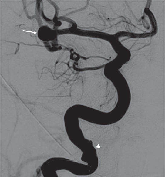
- A 64-year-old woman with a ruptured wide-necked bifurcation aneurysm of the right middle cerebral artery who presented with sudden onset severe headache. Digital subtraction angiography image taken during right internal carotid artery contrast injection showing a wide-necked right middle cerebral artery bifurcation aneurysm (arrow). Note mild irregularity of the distal cervical segment of internal carotid artery due to vasospasm (arrowhead).
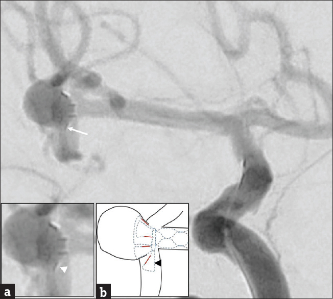
- A 64-year-old woman with a ruptured wide-necked bifurcation aneurysm of the right middle cerebral artery who presented with sudden onset severe headache. Intraprocedural digital subtraction angiography during right internal carotid artery injection after deploying the pCONus device showing one petal of the device (arrow) prolapsed out of the aneurysm neck into the inferior division of middle cerebral artery. Inset (a) magnified image of the aneurysm with the deployed pCONus device, the prolapsed petal shown by arrowhead. Inset (b) schematic diagram showing approximate outlines of the aneurysm and parent vessels (black outline), the four radiopaque markers of the petals (red lines) and the position of the distal petals and stent which are not radiopaque, hence not visible in angiogram. (Blue-colored dashed shapes); the prolapsed petal is indicated by a black arrowhead.
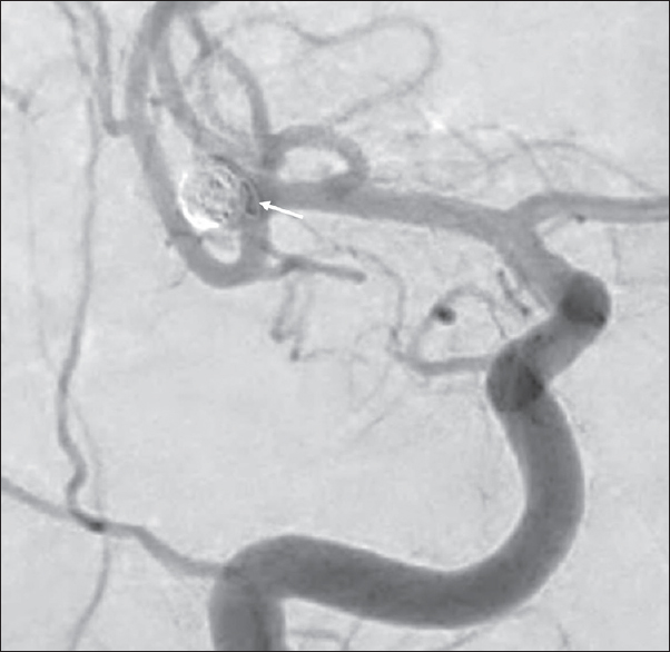
- A 64-year-old woman with a ruptured wide-necked bifurcation aneurysm of the right middle cerebral artery who presented with sudden onset severe headache. Postprocedural digital subtraction angiography during right carotid artery injection showing minimal filling of the aneurysm, patent middle cerebral artery branches, and prolapse of a coil loop into the middle cerebral artery (arrow).
Follow-up angiography done at 3 months showed minimal residual filling and stable aneurysm size. There was complete occlusion on follow up angiography performed 6 months later and the dissection in the internal carotid artery had healed without luminal compromise [Figure 4].
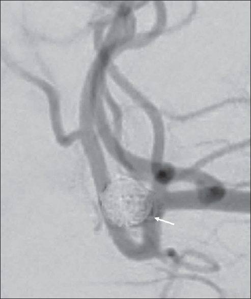
- A 64-year-old woman with a ruptured wide-necked bifurcation aneurysm of the right middle cerebral artery who presented with sudden onset severe headache. Digital subtraction angiography during right internal carotid artery contrast injection, performed during the 6th month follow-up visit showing occlusion of aneurysm, persistence of prolapsed coil loop (arrow), and normal flow in the middle cerebral artery and its branches.
DISCUSSION
Wide necked bifurcation aneurysms have always posed a challenge for endovascular treatment. Various strategies available for endovascular treatment of bifurcation aneurysms include coiling using 3D coils, balloon-assisted coiling using a single complaint or two balloons, stent-assisted coiling with two stents in Y or T configuration or single stent using “shelf” technique or waffle cone technique, barrel stent-assisted coiling, intraaneurysmal flow diversion using WEB device, and pCONus-assisted coiling.[13567] TriSpan and PulseRider devices which are designed specifically for wide necked bifurcation aneurysms are not widely used.[4] 3D coils have their limitations if the aneurysm is shallow, where even their shape alone is not enough to prevent prolapse into the parent vessel.[5] Balloon- or stent-assisted techniques require cannulation of the efferent vessels from the neck region of the aneurysm which is many a time technically very challenging.[15] The technique of cannulating an efferent vessel by creating of loop in the aneurysm and straightening the loop either with the “wire anchor loop traction” method or using a balloon, stent or a coil for anchoring is hazardous due to traction forces exerted on the aneurysm wall, especially if ruptured.[18910] Simultaneous placement of multiple devices and catheters as required in balloon- or stent-assisted coiling may cause hemodynamic compromise resulting in cerebrovascular events.[5] In addition, balloons have a propensity to cause dissection if overinflated and there is a possibility of coil prolapse on balloon deflation.[511] Placement of double balloon or double stents for coiling of wide-necked bifurcation aneurysms is laden with significant complication rates.[1112]
Deployment of a stent with its distal end within an aneurysm instead of bridging the neck is called the “waffle cone technique” and has been successfully applied using Neuroform, Enterprise, and Solitaire AB stents.[3131415] The pCONus device is a stent exclusively made for this purpose.[16] The pCONus device is a nitinol stent with distal radially flared four loops (petals) which are to be positioned at the base of the aneurysm and an additional six polyamide fibers at the distal diameter of the stent forming a barrier preventing coil prolapse into the parent vessels. The device is compatible with a 0.021″ microcatheter, is retrievable even after complete deployment, and is detached electrolytically. pCONus-assisted coiling is technically easier and superior to double stent-assisted coiling since there is no necessity to cannulate the efferent vessels and there is less intravascular metal placement.[5] There is, however, an argument that there may be a detrimental effect on the hemodynamics due to flow diversion toward the aneurysm.[1718] A study on intraaneurysmal flow kinetics using DSA optical flow imaging after deployment of pCONus device has shown no adverse increase in aneurysmal flow; on the contrary, it has shown a minimal reduction.[16] An added advantage of the device is that, in the case of aneurysm regrowth, the petals still remain at the neck of the aneurysm and facilitate coil retention within the aneurysm.[5]
The usage of pCONus device for ruptured aneurysms requires dual antiplatelets which might confer additional risk to the procedure, however, has been used successfully in multiple studies.[145] Thromboembolic complications, both transient (5%–20%) and permanent (2.5%–10.5%) have been documented in various studies.[1451920] Failed detachment of a pCONus device has been reported, where the device was retrieved without re-sheathing into the microcatheter resulting in a nonflow-limiting dissection.[1] Prolapse of a petal of the pCONus device can occur when the device sizing is improper (choosing smaller crown size than appropriate), if the aneurysm is eccentric to the parent vessel, or if excessive traction is applied to the pCONus device after it is deployed at the aneurysm neck. The pCONus device is compatible and has been used with a 0.021″ microcatheter. Un-re-sheath-ability of a deployed pCONus device is of serious concern in case the device is misaligned with one or more of its distal petals prolapsing into the parent vessels. In such cases, aneurysm can be coiled without an additional device if the first deployed 3D coil is supported within the aneurysm optimally by the device; if not, an additional balloon or a stent placed within the branch harboring the prolapsed petal can be used to support coil retention within the aneurysm. Metal coverage of pCONus of <5%[1] would not cause any difficulty in placement of an additional stent or a balloon, and there is a description of placement of two stents through a pCONus device for a successful treatment of a basilar artery aneurysm.[5] Removal of the misaligned pCONus device in its deployed state using gentle traction is another option, as has been described once in a case of failed detachment of the device by Fischer et al.,[1] but was not performed in this case fearing the risk of endothelial damage to the aneurysm neck and parent vessel which might result in a dissection, which in unfortunate circumstances could be flow limiting or occluding. Probably, the usage of a 0.027″ inner diameter microcatheter is likely to avoid this complication. To the best of our knowledge, un-re-sheath-ability of a misaligned pCONus device has not been described yet in literature; and such a complication is of serious concern if the misaligned device does not support coil retention within the aneurysm.
Declaration of patient consent
The authors certify that they have obtained all appropriate patient consent forms. In the form the patient(s) has/have given his/her/their consent for his/her/their images and other clinical information to be reported in the journal. The patients understand that their names and initials will not be published and due efforts will be made to conceal their identity, but anonymity cannot be guaranteed.
Financial support and sponsorship
Nil.
Conflicts of interest
There are no conflicts of interest.
Available FREE in open access from: http://www.clinicalimagingscience.org/text.asp?2017/7/1/41/217047
REFERENCES
- Single-center experience in the endovascular treatment of wide-necked intracranial aneurysms with a bridging intra-/extra-aneurysm implant (pCONus) J Neurointerv Surg. 2016;8:1186-91.
- [Google Scholar]
- Waffle Y technique: PCONus for tandem bifurcation aneurysms of the middle cerebral artery. J Neurointerv Surg. 2014;6:e51.
- [Google Scholar]
- “Waffle cone” technique for intra/extra-aneurysmal stent placement for the treatment of complex and wide-necked bifurcation aneurysm. Interv Neuroradiol. 2008;14(Suppl 2):49-52.
- [Google Scholar]
- PCONus device for the endovascular treatment of wide-neck middle cerebral artery aneurysms. AJNR Am J Neuroradiol. 2015;36:1735-40.
- [Google Scholar]
- Coil occlusion of wide-neck bifurcation aneurysms assisted by a novel intra- to extra-aneurysmatic neck-bridging device (pCONus): Initial experience. AJNR Am J Neuroradiol. 2014;35:965-71.
- [Google Scholar]
- LVIS Jr “shelf” technique: An alternative to Y stent-assisted aneurysm coiling. J. Neurointerv Surg 2016 pii: neurintsurg-2015-012246
- [Google Scholar]
- WEB device for endovascular treatment of wide-neck bifurcation aneurysms. AJNR Am J Neuroradiol. 2013;34:1209-14.
- [Google Scholar]
- The wire anchor loop traction (WALT) maneuver. BMJ Case Rep 2015 2015 pii: Bcr2014011604
- [Google Scholar]
- The balloon anchor technique: A novel technique for distal access through a giant aneurysm. J Neurointerv Surg. 2010;2:363-7.
- [Google Scholar]
- The stent anchor technique for distal access through a large or giant aneurysm. J Neurointerv Surg. 2013;5:e24.
- [Google Scholar]
- Safety and efficacy of balloon remodeling technique during endovascular treatment of intracranial aneurysms: Critical review of the literature. AJNR Am J Neuroradiol. 2012;33:12-5.
- [Google Scholar]
- Stent-supported aneurysm coiling: A literature survey of treatment and follow-up. AJNR Am J Neuroradiol. 2012;33:159-63.
- [Google Scholar]
- Waffle-cone technique using solitaire AB stent. J Korean Neurosurg Soc. 2012;51:222-6.
- [Google Scholar]
- Intra/extra-aneurysmal stent placement for management of complex and wide-necked-bifurcation aneurysms: Eight cases using the waffle cone technique. Neurosurgery. 2006;58:ONS.
- [Google Scholar]
- Stent application for the treatment of cerebral aneurysms. Neurointervention. 2011;6:53-70.
- [Google Scholar]
- Intra-aneurysmal hemodynamics: Evaluation of pCONus and pCANvas bifurcation aneurysm devices using DSA optical flow imaging. J Neurointerv Surg. 2016;8:1197-201.
- [Google Scholar]
- Stent-assisted coiling of intracranial bifurcation aneurysms leads to immediate and delayed intracranial vascular angle remodeling. AJNR Am J Neuroradiol. 2012;33:649-54.
- [Google Scholar]
- Waffle-cone technique with Solitaire™ AB remodeling device: Endovascular treatment of highly selected complex cerebral aneurysms. Neuroradiology. 2011;53:961-72.
- [Google Scholar]
- The pCONus device for the endovascular treatment of wide neck bifurcation aneurysms. J Neurointerv Surg. 2016;8:940-4.
- [Google Scholar]
- The pCONus device in treatment of wide.necked aneurysms: Technical and midterm clinical and angiographic results. 2016. Clin Neuroradiol. :1-8. Available from: https://www.link.springer.com/article/10.1007%2Fs00062-016-0542-z
- [Google Scholar]


