
Translate this page into:
Accuracy of Computerized Vertical Measurements on Digital Orthopantomographs: Posterior Mandibular Region
-
Received: ,
Accepted: ,
This is an open-access article distributed under the terms of the Creative Commons Attribution License, which permits unrestricted use, distribution, and reproduction in any medium, provided the original author and source are credited.
This article was originally published by Medknow Publications & Media Pvt Ltd and was migrated to Scientific Scholar after the change of Publisher.
Abstract
Objectives:
Orthopantomographs are commonly used for diagnosis in clinical dentistry. Although the manufacturers claim a constant magnification effect, the reliability of measuring dimensions on the panoramic radiographs is not clear. The aim of this study was to evaluate the accuracy of measuring vertical dimensions in the posterior mandibular area on digital orthopantomographs.
Materials and Methods:
A retrospective survey of 20 orthopantomographs with unrestored implants (only with cover screw) in the mandibular posterior region (molars and premolars) was conducted. All radiographs were taken using the same machine by skilled technicians. Two examiners were asked to measure the vertical dimension of the implants seen on the radiographs viewed using two differently sized display screens. Inter-examiner and intra-examiner reliability tests were performed. Differences between the measured length and the actual length using each screen type were compared.
Results:
High coefficients of reliability were observed on intra- and inter-examiner correlation. The overall reliability of measuring the vertical dimensions of implants between both examiners for the large screen and the small screen were 97.4% (Cronbach's alpha 0.993) and 94.0% (Cronbach's alpha 0.984), respectively. There were no significant differences between the errors seen with either the large screen or the small screen, when each of them was compared to the original length (P = 0.146).
Conclusion:
This study shows that vertical dimensions in the posterior mandibular region (molar and premolars) can be reliably measured on an orthopantomograph using a calibrated machine and special software.
Keywords
Digital panoramic radiograph
implant length
orthopantomographs
posterior mandible
vertical measurements

INTRODUCTION

After W. C. von Roentgen discovered X-rays in 1896, a German dentist, O. Walkhoff, used radiographs for dental diagnosis.[1] At the beginning of the last century, attempts were made to image the whole jaw with intraoral radiography. Panoramic technique developed to image the teeth and jaws became an essential element in oral radiology.[2]
Orthopantomography (OPT) became a very popular and widely accepted technique of dental radiography. It is a curved plane tomographic radiographic technique used to depict on a single image the body of the mandible and maxilla, the lower part of the maxillary sinuses, and the temporomandibular joints.[3] OPTs have a wide variety of uses, including the screening of patients during dental treatment for evidence of cysts, impacted teeth, foreign bodies, and neoplasms.[45] OPTs are also used to evaluate pathological situations related to the temporomandibular joints and maxillary sinuses and to evaluate mandibular fractures. In addition, OPTs offer information about the locations of important anatomic structures in the orofacial region which are needed for dental implant planning.[67]
The evolution of more precise three-dimensional techniques such as conventional computed tomography (CT), cone beam computed tomography (CBCT), and magnetic resonance imaging has improved oral diagnosis and implant treatment planning by virtue of their higher precision.[89] In comparison with CT and other expensive precision radiographs, OPT is a simple, low-cost imaging modality with patients being exposed to relatively low dose of radiation.[101112] The effective doses may vary significantly among CBCT machines; however, when compared to medical CT, CBCT is considered to be a low-dose technique for use in dental implant procedure.[131415] The effective dose from CBCT examinations could range from 13 to 479 μSv with different commercially available CBCT machines, while the effective dose from one OPT is approximately 10–14 μSv.[13] On the other hand, the exposure from a maxillomandibular medical CT ranges from 474 to 1160 μSv.[16]
Digital panoramic machines are not significantly different from conventional panoramic units; thus, it has been suggested that the degree of vertical magnification due to projection geometry is similar for digital and conventional rotational OPTs.[9] Calibrated software-based tools are used to overcome the magnification when measuring vertical dimensions on a digital OPT; the measurements will be more accurate when the magnification of the radiograph is closer to the estimated magnification predetermined by the manufacturer. In 1986, Larheim and Svanaes[17] investigated the precision of measurements of mandibular linear dimensions in panoramic radiographs and showed that the variability of vertical measurements made from repeated panoramic radiographs was small when patients were properly positioned in the panoramic machine. They reported that the highest reliability was obtained when the same radiographer adjusted the head position and made both exposures.[17]
There are a large number of panoramic X-ray machines available from various manufacturers, and the magnification factor varies from one manufacturer to another. This variation results in differences in magnification and in the amounts of distortion and displacement of structures.[1819] Other factors such as the skills of the examiner and the position of the patient's head may also reduce the accuracy of the OPT.[20] The aim of this study is to evaluate the accuracy of measuring vertical dimensions in the posterior mandibular area on a single digital OPT machine, and to determine the margin of error of measurements from OPTs taken in regular daily practice. The study also evaluated the possible influence of the size of the display screen on the accuracy of measurements.
MATERIALS AND METHODS
A retrospective review of the files of all implant patients treated at Alpha Clinic, a private clinic limited to periodontics and dental implants (Ramallah, Palestine), during the years 2010–2013 was conducted. After exclusion of non-suitable images, the selection process yielded a total of 20 digital panoramic radiographs taken using the same radiographic machine. These radiographs were taken during the process of treatment for various reasons; none were taken for the purpose of this study. A total of 27 implants inserted in the posterior segment of the mandible were included for evaluation. Eleven (60%) implants were placed in the premolar region and 16 (40%) in the molar region. Seven of the selected panoramic radiographs included more than one implant, while six implants were evaluated in two different panoramic radiographs at different time points. These images were taken from the records of 14 patients (6 males and 8 females; age 20–51 years, mean age 34 years) [Table 1].

All implants in the OPTs selected were placed by a single surgeon (M. A.) who decided the width and length (L) of the implants based on pre-operative CBCT images used for treatment planning. The location of dental implants for a partial edentulous ridge was determined clinically, considering the adjacent and opposing teeth. The included implants were of different systems, models, and dimensions. The lengths of dental implants used in the obtained OPTs ranged between 8 and 13 mm [Table 1].
Radiograph inclusion criteria
-
Only panoramic radiographs with implants in the molar and premolar mandibular regions were included in this study.
-
All panoramic radiographs used in this study were required to be taken using one single radiographic machine: KODAK 9000 3D® CBCT (Carestream Health, Inc., Marne-la-Vallée, France). These images were taken as direct OPTs and not as CBCT reconstructions.
-
The panoramic radiograph included had to clearly show the inferior alveolar nerve, the mental foramen, the nasal floor, and the maxillary sinus floor. The clarity of images was subjectively determined by a single dentist (A. AG.) who is experienced in interpreting panoramic radiographs.
-
To avoid measurement mistakes, only root form titanium dental implants covered by covering screw (without healing cap, temporary restoration, or crown) were included in this study.
Radiography details
All digital panoramic radiographs were taken at Qirrish Center for Oral Radiology (Ramallah, Palestine) which has one radiographic device for panoramic X-ray (KODAK 9000 3D® CBCT). Digital panoramic radiographs were taken under everyday conditions by two skilled technicians according to the manufacturer's specified posit ion for the patient's head, but did not follow a strict, standardized protocol to ensure the patient's precise head position for any research purpose. Images were delivered through a compact disk to our office to be stored on our computer for evaluation using a specialized computer software (Dental Imaging Software DIS Patient File 6.13.0.24, Carestream Health, Inc., 2013) which is designed specifically for storage and interpretation of the digital data received from the X-ray machine (KODAK 9000 3D® ).
Measurements
The vertical length of each included implant shown on digital panoramic radiographs was measured by two independent examiners, who were blind to the actual readings and did not participate in the treatment of the patients. Examiner (A) is a pedodontist; the other examiner (B) is a general dental practitioner with 2 years of experience in dentistry. Neither of them was aware of the actual size of the implants used nor were they familiar with the implant systems used at our clinic. Each examiner had to examine randomly the X-rays at two different sessions with 1 week interval. At each session, the examiner had to examine each X-ray using a different display screen attached to the computer; a large screen (LS) of 42 inches 50–60 Hz 1280 × 768 pixels at 60 Hz (Philips 42PFL3606H/12; 2011) and a small screen (SS) of 19 inches, 50–60 Hz 1440 × 900 pixels at 60 Hz (HP NK570A; 2009).
Each examiner was requested to use a mouse-driven pointer to select the most apical point and the most coronal point of each implant on the computer software, which will automatically give the estimated distance between these two selected points to the nearest tenth of a millimeter. This reading which represents the measured length of the implant was recorded by the examiner [Figure 1].
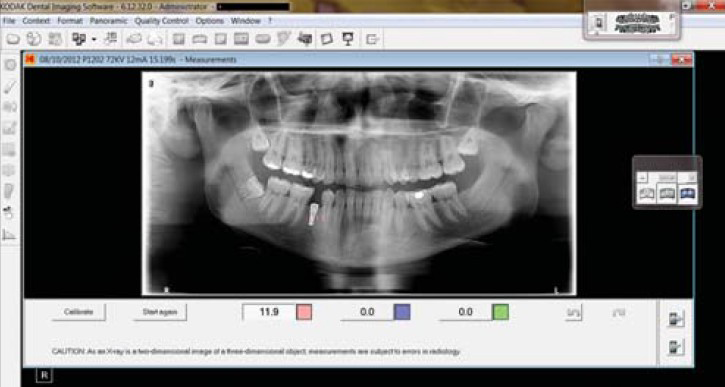
- Measurement using a computer software tool to determine the vertical dimension of an implant placed in the lower jaw. The radiograph shows the calculated length between the two points selected by the examiner using the computer's mouse.
Statistical analysis
Data analysis included tabulation of descriptive statistics. Reliability analysis was assessed using intra-class correlation coefficients and Cronbach's alpha statistic. For each individual examiner, intra-examiner reliability statistics for LS and SS were drawn. Inter-examiner reliability statistics for each type of measured LS and SS were also drawn. This reading which represents the measured length Mean LS, mean SS, and L measures were compared using Related Samples Friedman's two-way analysis of variance (ANOVA). The error between L and measured implant length (for LS and SS) was computed (dLS and dSS for each examiner, respectively). Mean error, dLS, and dSS values were computed and compared using Related Samples Wilcoxon's signed rank test.
RESULTS
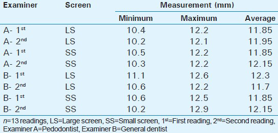
Of all the 20 selected OPTs, none was excluded due to lack of clarity or for having major distortions. Descriptive demographic data about the actual size and location of the implants is presented in Table 1. The actual diameters according to the files of the patients ranged between 3.2 and 5.0 mm. The actual lengths according to patient records ranged between 8 and 13 mm; the most frequently present length was 11.5 mm which was present in 13 radiographs. The minimal and maximal readings by each examiner using the different screens of the 11.5 mm implants at each reading are shown in Table 2.
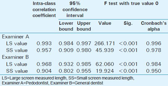
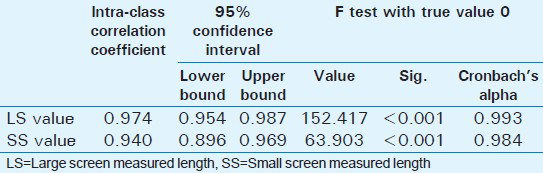
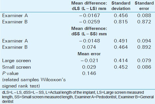
The intra-examiner reliability (intra-class coefficient measure) was 99.3% and 95.7% for examiner A on LS and SS, respectively [Table 3]. The intra-examiner reliability (intra-class coefficient measure) was 96.8% and 90.4% for examiner B on LS and SS, respectively [Table 3]. The inter-examiner reliability (intra-class coefficient measure) for LS was 97.4% for both examiners [Table 4]. The inter-examiner reliability (intra-class coefficient measure) for SS was 94. 0% for both examiners [Table 4].
For LS, the mean error compared to L was − 0.21 mm (SD 0.414 mm) with both raters [Table 5]. For SS, the mean error compared to L was 0.29 mm (SD 0.452 mm) with both raters [Table 5]. There were no significant differences between L, mean LS, and mean SS measures (P = 0.891, Related Samples Friedman's ANOVA). There was no significant difference between the mean errors via LS and SS measures when Related Samples Wilcoxon's signed rank test was used to compare the median of differences (P = 0.146).
DISCUSSION
The examiners who measured the vertical dimensions of implants on the digital screens were unaware of the details of the patients and implants. Although both of them are dentists, they have very minimal experience in dental implantology. They were asked not to try to figure out what were the possible lengths of implants, so that their measurements would not be biased to a certain length. Although the dental implants in OPTs were of various brands, no information was given to the examiners about which types of implants we use in our clinic, in order to avoid any possible bias by the examiner who might try to guess the length of implant and influence the measurements. Each examiner was asked to do the measurements twice on different days with at least 1 week interval to measure the intra-examiner reliability. The reliability for repeated measurements was high for both examiners, whether they were using the LS or SS.
Accuracy of an OPT is influenced by patient's head position, the observer's experience, and accuracy.[20] All these factors could be eliminated to the minimum if the radiographer was knowing that the OPT would be analyzed for research purposes; thus, the retrospective design of this study gives more realistic results. The calibration of the machine is another factor that can impact the accuracy and reproducibility obtained from digital panoramic radiographs.[9181921] For this reason, we used all radiographs taken by the same machine to eliminate calibration variations between different devices. Two skilled radiographers work at the radiology center where the OPTs were taken; either one of them could have taken any of the radiographs we received. There is no information of who was the radiographer of each OPT, because they do not write such details in their routine records. Not revealing this data makes our study more realistic and reduces the bias, because it has been reported in a previous study that the reproducibility of OPTs could be different between two different radiographers using the same machine.[17]
Two different screen sizes were utilized to determine if this factor may influence the accuracy of measuring the vertical length; however, the inter-class coefficient showed that there were no significant differences in measurements according to screen sizes [Table 4].
High accuracy was shown in this study [Table 5], where the maximal error in measurements never exceeded the precautionary safety margin of 2 mm between the drilling position for implant site preparation and any vital anatomic landmark.[2223]
In the 1980s, the first revolution came with the introduction of digital dental radiography in dentistry. A second milestone was reached in the 1990s with the introduction of computerized software applications for two- and three-dimensional diagnostics which could be efficiently used for presurgical planning.[242526] The imaging technology is still a promising field in surgical planning for dental implants. Probably the use of three-dimensional images may ensure better precision in implant techniques.
Image magnification and lack of cross-sectional information are the major drawbacks of the image modality of OPTs for implant surgery planning.[27] Thus, it is not possible to confirm that the dimensions of structures shown on OPTs correspond to the real dimensions of the structures.[28] Different authors have reported vertical magnification of OPTs in the posterior mandibular area to be constant between 125 and 130%.[122930] Insignificantly slightly higher magnification was observed in the lower premolar area compared to molar area.[1230] Vazquez et al., suggested that the implant length measures could be used to evaluate the vertical magnification factor even when the patient's head position was not strictly standardized before exposure and when measurements were taken by observers with different skill levels and experience.[23] These results are consistent with this study which was performed retrospectively in a daily practice environment.
Other studies have both confirmed the reliability of vertical dimensions on OPTs and shown that horizontal assessments are unreliable, especially in the anterior regions.[2331] Distortions in the anterior area could be caused by the fact that the curvature level of the jaw is different in each individual and can be influenced by patient position during imaging.[32] A tendency to greater enlargements of measurements on OPTs has been observed in the maxilla compared to the mandible.[12]
To rely on the vertical dimension measured on an OPT for implant planning, Philip Worthington[22] has suggested a simple formula which considers magnification of the radiograph, a “safety zone” margin (1–2 mm), and the “useless” thin crestal bone. He recommended performing careful measurement and using the correct magnification factor. He suggested that nerve injury should be included in the informed consent, and both radiograph and calculations should be kept in patient's chart as evidence of meticulous patient care. Other important local factors that might influence the accuracy of measuring the vertical height of available bone in the posterior mandibular area using an OPT are the bucco-lingual position of the inferior alveolar canal and the bucco-lingual position of crestal peak of alveolar bone. The bucco-lingual positions of these landmarks may result in false estimations of the height of available alveolar bone.[2227]
Limitations
The limitation of this study is the small number of radiographs used in this retrospective study. A study involving a greater number of radiographs will give more accurate results.
CONCLUSION
Within the limitations of this study, it seems that digital OPT is a reliable and safe imaging modality to evaluate the vertical dimension of the alveolar bone in posterior mandibular (molar and premolar) regions if a well-calibrated machine with a specialized measuring software is used. Using a high-quality screen is important; however, there were no significant differences between the measurements obtained by different screen sizes used in this study. A larger scale study needs to be conducted with more radiographs and examiners to confirm these results.
Available FREE in open access from: http://www.clinicalimagingscience.org/text.asp?2014/4/2/7/148274
Source of Support: Nil
Conflict of Interest: None declared.
REFERENCES
- Dental cone beam computed tomography: Justification for use in planning oral implant placement. Periodontol 2000. 2014;66:203-13.
- [Google Scholar]
- A survey of the radiographic practices of general dentists for edentulous patients. Oral Surg Oral Med Oral Pathol Oral Radiol Endod. 1995;80:365-8.
- [Google Scholar]
- Location of the mandibular foramen in panoramic radiographs. Oral Surg Oral Med Oral Pathol. 1994;78:662-9.
- [Google Scholar]
- Patient assessment and diagnosis in implant treatment. Aust Dent J. 2008;53(Suppl 1):S3-10.
- [Google Scholar]
- Guidelines for radiologic examinations: Do we have all the answers yet? Oral Surg Oral Med Oral Pathol Oral Radiol Endod. 1997;83:523-4.
- [Google Scholar]
- Accuracy of vertical height measurements on direct digital panoramic radiographs using posterior mandibular implants and metal balls as reference objects. Dentomaxillofac Radiol. 2013;42:20110429.
- [Google Scholar]
- Diagnostic value of dental CT (DentaScan) in dental implant. Chungbuk Med J. 1998;8:11-19.
- [Google Scholar]
- Efficacy of panoramic radiographs in the preoperative planning of posterior mandibular implants: A prospective clinical study of 1527 consecutively treated patients. Clin Oral Implants Res. 2008;19:81-5.
- [Google Scholar]
- Magnification rate of digital panoramic radiographs and its effectiveness for pre-operative assessment of dental implants. Dentomaxillofac Radiol. 2011;40:76-83.
- [Google Scholar]
- Dosimetry of 3 CBCT devices for oral and maxillofacial radiology: CB Mercuray, NewTom 3G and i-CAT. Dentomaxillofac Radiol. 2006;35:219-26.
- [Google Scholar]
- Absorbed and effective doses from cone beam volumetric imaging for implant planning. Dentomaxillofac Radiol. 2009;38:79-85.
- [Google Scholar]
- Effective radiation dose of ProMax 3D cone-beam computerized tomography scanner with different dental protocols. Oral Surg Oral Med Oral Pathol Oral Radiol Endod. 2010;110:770-6.
- [Google Scholar]
- Comparison between effective radiation dose of CBCT and MSCT scanners for dentomaxillofacial applications. Eur J Radiol. 2009;71:461-8.
- [Google Scholar]
- Reprodicibility of rotational panoramic radiography: Mandibular linear dimensions and angles. Am J Orthod Dentofacial Orthop. 1986;90:45-51.
- [Google Scholar]
- Comparison of linear dimensions and angular measurements on panoramic images taken with two machines. J Dent Res Dent Clin Dent Prospects. 2009;3:7-10.
- [Google Scholar]
- A study of the focal troughs of three panoramic dental X-ray machines, I: The area of sharpness. Oral Surg Oral Med Oral Pathol. 1975;39:318-28.
- [Google Scholar]
- The quality of panoramic radiographs in a sample of general dental practices. Br Dent J. 1999;186:630-3.
- [Google Scholar]
- Condylar height on panoramic radiographs. A methodological study with a clinical application. Acta Odontol Scand. 1994;52:43-50.
- [Google Scholar]
- Injury to the inferior alveolar nerve during implant placement: A formula for protection of the patient and clinician. Int J Oral Maxillofac Implants. 2004;19:731-4.
- [Google Scholar]
- Reliability of the vertical magnification factor on panoramic radiographs: Clinical implications for posterior mandibular implants. Clin Oral Implants Res. 2011;22:1420-5.
- [Google Scholar]
- Predictability of a three-dimensional planning system for oral implant surgery. Dentomaxillofac Radiol. 1999;28:105-11.
- [Google Scholar]
- Computer-assisted planning of oral implant surgery: A three dimensional approach. Int J Oral Maxillofac Implants. 1996;11:806-10.
- [Google Scholar]
- Computer-assisted planning of oral implant surgery. An approach using virtual reality. Stud Health Technol Inform. 1996;29:423-34.
- [Google Scholar]
- The effects of location of alveolar crest on the vertical bone heights on panoramic radiographs. Dentomaxillofac Radiol. 2012;41:117-21.
- [Google Scholar]
- Evaluation of the precision of dimensional measurements of the mandible on panoramic radiographs. Oral Surg Oral Med Oral Pathol Oral Radiol Endod. 1998;86:242-8.
- [Google Scholar]
- Study on the necessity for cross-section imaging of the posterior mandible for treatment planning of standard cases in implant dentistry. Clin Oral Implants Res. 2004;15:490-7.
- [Google Scholar]
- The evaluation of digital panoramic radiographs taken for implant dentistry in daily practice. Med Oral Patol Oral Cir Bucal. 2010;15:e663-6.
- [Google Scholar]
- Image distortion in rotational panoramic radiography. II. Vertical distances. Acta Radiol Diagn (Stockh). 1981;22:449-55.
- [Google Scholar]
- Imaging technique selection for the pre-operative planning of oral implants: A review of the literature. Clin Implant Dent Relat Res. 2002;4:156-72.
- [Google Scholar]


